| Russian language (monolingual) [PRO] Medical — Medical (general) |
||||||
|---|---|---|---|---|---|---|
|
|
|||||
|
Selected response from: Yana Onikiychuk |
|||||
|
Grading comment 4 KudoZ points were awarded for this answer |


| SUMMARY OF ALL EXPLANATIONS PROVIDED | ||||
|---|---|---|---|---|
| 5 +6 | см. |
|
||
| 4 +5 | двенадцатиперстная кишка |
|
||
| 5 +3 | Е (не Ё) |
|
||
| 4 +4 | двенадцатипЕрстная |
|
||
| 5 +3 | просто е |
|
||
| 4 +3 | двенадцатиперстная кишка |
|
| Summary of reference entries provided | |||
|---|---|---|---|
|
Answers
2 mins confidence: ![]() peer agreement (net): +3
peer agreement (net): +3
2 mins confidence: ![]() peer agreement (net): +6
peer agreement (net): +6
| duodenum
см.
Explanation: |
Yana Onikiychuk
Russian Federation Local time: 23:56 Specializes in field Native speaker of: Russian PRO pts in category: 4
|
||||
|
|
|||||
| Login to enter a peer comment (or grade) |
2 mins confidence: ![]()
![]() peer agreement (net): +4
peer agreement (net): +4
3 mins confidence: ![]()
![]() peer agreement (net): +3
peer agreement (net): +3
3 mins confidence: ![]() peer agreement (net): +3
peer agreement (net): +3
| duodenum
Е (не Ё)
Explanation: ————————————————— орфографический словарь, 106000 слов |
Alexander Ryshow
Belarus Local time: 00:56 Works in field Native speaker of:  Russian Russian
|
|
|
|
| Login to enter a peer comment (or grade) |

2 mins confidence: ![]()
![]() peer agreement (net): +5
peer agreement (net): +5
| duodenum
двенадцатиперстная кишка
Explanation: ————————————————— В любом случае, при наборе в gooogle «двенадцатипёрстная» поисковик все равно выдает букву Е. |
Alieksei Seniukovich
Belarus Local time: 00:56 Specializes in field Native speaker of: Russian PRO pts in category: 4
|
||||
|
|
|||||
| Login to enter a peer comment (or grade) |

Login or register (free and only takes a few minutes) to participate in this question.
You will also have access to many other tools and opportunities designed for those who have language-related jobs
(or are passionate about them). Participation is free and the site has a strict confidentiality policy.
Смотреть что такое ДВЕНАДЦАТИПЕРСТНАЯ КИШКА в других словарях:
ДВЕНАДЦАТИПЕРСТНАЯ КИШКА
(duodenum) — первый отдел тонкой кишки, названный так потому, что длина ее у человека равняется приблизительно 12 поперечникам пальца, всего около 30 с… смотреть
ДВЕНАДЦАТИПЕРСТНАЯ КИШКА
начальный отрезок тонкой кишки (от выходного отверстия желудка до тощей кишки, (см. рис.), четко обособленный у человека и млекопитающих животн… смотреть
ДВЕНАДЦАТИПЕРСТНАЯ КИШКА
Двенадцатиперстная кишка (duodenum) — первый отдел тонкой кишки, названный так потому, что длина ее у человека равняется приблизительно 12 поперечникам пальца, всего около 30 см. Начинаясь от привратника, места выхода из желудка, Д. кишка идет сперва направо и назад, загибаясь затем вниз впереди правой почки. В нисходящей части она выстлана брюшиной лишь спереди, тогда как задняя сторона ее рыхлой соединительной тканью прикрепляется к правой почке и к позвоночнику. Далее Д. кишка вновь поворачивает вперед и идет в поперечном направлении влево, впереди нижней полой вены и аорты; эта поперечная часть ее слегка согнута в виде дуги, выпуклость которой направлена вниз и вправо; затем Д. кишка переходит в тощую (jejunum). В общем Д. кишка имеет, следовательно, форму подковы, выпуклость которой направлена вправо, назад и вниз; нижнее поперечное колено этой подковы длинное, верхнее — очень короткое. Между обоими коленами, в вогнутости подковы, лежит головка поджелудочной железы, плотно прикрепленной в этом месте к Д. кишке. В слизистой оболочке Д. кишки, кроме рассеянных по всей тонкой кишке Либеркюновых желез, лежат еще особые Бруннеровы железы — маленькие, гроздевидные железки, лежащие частью в самой слизистой оболочке, частью под ней. В Д. кишку открываются выводные протоки печени и поджелудочной железы. <i> В. Ф. </i><br><br><br>… смотреть
ДВЕНАДЦАТИПЕРСТНАЯ КИШКА
(duodenum)начальный отдел тонкой кишки, расположенный между желудком и тощей кишкой.
Спереди Д. к. прикрывают желудок, правая доля печени и брыжейка п… смотреть
ДВЕНАДЦАТИПЕРСТНАЯ КИШКА
Двенадцатиперстная кишка, duodenum, начинается под печенью на уровне тела XII грудного или I поясничного позвонка, справа от позвоночного столба. На… смотреть
ДВЕНАДЦАТИПЕРСТНАЯ КИШКА
ДВЕНАДЦАТИПЕРСТНАЯ КИШКА
(duodenum), начальный отрезок тонкой кишки (от выходного отверстия желудка до тощей кишки), чётко обособленный у птиц и млеко… смотреть
ДВЕНАДЦАТИПЕРСТНАЯ КИШКА
1) Орфографическая запись слова: двенадцатиперстная кишка2) Ударение в слове: двенадцатип`ерстная кишк`а3) Деление слова на слоги (перенос слова): двен… смотреть
ДВЕНАДЦАТИПЕРСТНАЯ КИШКА
нач. отдел тонкой кишки (от выходного отверстия желудка до тощей кишки), хорошо обособленный у птиц, млекопитающих ж-ных и человека. Длина Д. к. челове… смотреть
ДВЕНАДЦАТИПЕРСТНАЯ КИШКА
ДВЕНАДЦАТИПЕРСТНАЯ КИШКА, начальный отдел тонкой кишки (от выходного отверствия желудка до тощей кишки), хорошо обособленный у птиц, млекопитающих животных и человека. Длина двенадцатиперстной кишки человека равна поперечникам 12 пальцев (отсюда название). Клетки слизистой оболочки двенадцатиперстной кишки вырабатывают кишечный сок и гормон секретин. В двенадцатиперстной кишке открываются проток поджелудочной железы и желчный проток. Основной вид патологии — язва.<br><br><br>… смотреть
ДВЕНАДЦАТИПЕРСТНАЯ КИШКА
ДВЕНАДЦАТИПЕРСТНАЯ КИШКА — начальный отдел тонкой кишки (от выходного отверствия желудка до тощей кишки), хорошо обособленный у птиц, млекопитающих животных и человека. Длина двенадцатиперстной кишки человека равна поперечникам 12 пальцев (отсюда название). Клетки слизистой оболочки двенадцатиперстной кишки вырабатывают кишечный сок и гормон секретин. В двенадцатиперстной кишке открываются проток поджелудочной железы и желчный проток. Основной вид патологии — язва.<br>… смотреть
ДВЕНАДЦАТИПЕРСТНАЯ КИШКА
ДВЕНАДЦАТИПЕРСТНАЯ КИШКА , начальный отдел тонкой кишки (от выходного отверствия желудка до тощей кишки), хорошо обособленный у птиц, млекопитающих животных и человека. Длина двенадцатиперстной кишки человека равна поперечникам 12 пальцев (отсюда название). Клетки слизистой оболочки двенадцатиперстной кишки вырабатывают кишечный сок и гормон секретин. В двенадцатиперстной кишке открываются проток поджелудочной железы и желчный проток. Основной вид патологии — язва…. смотреть
ДВЕНАДЦАТИПЕРСТНАЯ КИШКА
ДВЕНАДЦАТИПЕРСТНАЯ КИШКА, начальный отдел тонкой кишки (от выходного отверствия желудка до тощей кишки), хорошо обособленный у птиц, млекопитающих животных и человека. Длина двенадцатиперстной кишки человека равна поперечникам 12 пальцев (отсюда название). Клетки слизистой оболочки двенадцатиперстной кишки вырабатывают кишечный сок и гормон секретин. В двенадцатиперстной кишке открываются проток поджелудочной железы и желчный проток. Основной вид патологии — язва…. смотреть
ДВЕНАДЦАТИПЕРСТНАЯ КИШКА
— начальный отдел тонкой кишки (от выходногоотверствия желудка до тощей кишки), хорошо обособленный у птиц,млекопитающих животных и человека. Длина двенадцатиперстной кишки человекаравна поперечникам 12 пальцев (отсюда название). Клетки слизистой оболочкидвенадцатиперстной кишки вырабатывают кишечный сок и гормон секретин. Вдвенадцатиперстной кишке открываются проток поджелудочной железы и желчныйпроток. Основной вид патологии — язва…. смотреть
ДВЕНАДЦАТИПЕРСТНАЯ КИШКА
ДВЕНАДЦАТИПЕРСТНАЯ КИШКА, первый отдел тонкого КИШЕЧНИКА, имеющий вид подковы, часть ПИЩЕВАРИТЕЛЬНОЙ СИСТЕМЫ. От ЖЕЛУДКА ее отделяет круговая мышца — п… смотреть
ДВЕНАДЦАТИПЕРСТНАЯ КИШКА
Начальная форма — Двенадцатиперстная кишка, единственное число, женский род, именительный падеж, неодушевленное
двенадцатиперстная кишка
Правильное написание:
двенадцатипе́рстная кишка́
Рады помочь вам узнать, как пишется слово «двенадцатиперстная кишка».
Пишите и говорите правильно.
О словаре
Сайт создан на основе «Русского орфографического словаря», составленного Институтом русского языка имени В. В. Виноградова РАН. Объем второго издания, исправленного и дополненного, составляет около 180 тысяч слов, и существенно превосходит все предшествующие орфографические словари. Он является нормативным справочником, отражающим с возможной полнотой лексику русского языка начала 21 века и регламентирующим ее правописание.
From Wikipedia, the free encyclopedia
| Duodenum | |
|---|---|

Image of the gastrointestinal tract, with the duodenum highlighted. |
|
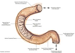
Diagram of the human duodenum with major parts labelled |
|
| Details | |
| Pronunciation | |
| Precursor | Foregut (1st and 2nd parts), Midgut (3rd and 4th part) |
| Part of | Small intestine |
| System | Digestive system |
| Artery | Inferior pancreaticoduodenal artery, Superior pancreaticoduodenal artery |
| Vein | Pancreaticoduodenal veins |
| Nerve | celiac ganglia, vagus[1] |
| Identifiers | |
| Latin | Intestinum duodenum |
| MeSH | D004386 |
| TA98 | A05.6.02.001 |
| TA2 | 2944 |
| FMA | 7206 |
| Anatomical terminology
[edit on Wikidata] |
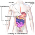
The duodenum is the first section of the small intestine[2] in most higher vertebrates, including mammals, reptiles, and birds. In mammals it may be the principal site for iron absorption.
The duodenum precedes the jejunum and ileum and is the shortest part of the small intestine.
In humans, the duodenum is a hollow jointed tube about 25–38 centimetres (10–15 inches) long connecting the stomach to the middle part of the small intestine.[3][4] It begins with the duodenal bulb and ends at the suspensory muscle of duodenum.[5] Duodenum can be divided into four parts: the first (superior), the second (descending), the third (horizontal) and the fourth (ascending) parts.[4]
Overview[edit]
The duodenum is the first section of the small intestine in most higher vertebrates, including mammals, reptiles, and birds. In fish, the divisions of the small intestine are not as clear, and the terms anterior intestine or proximal intestine may be used instead of duodenum.[6] In mammals the duodenum may be the principal site for iron absorption.[7]
In humans, the duodenum is a C-shaped hollow jointed tube, 25–38 centimetres (10–15 inches) in length, lying adjacent to the stomach (and connecting it to the small intestine). It is divided anatomically into four sections. The first part lies within the peritoneum but its other parts are retroperitoneal.[8]: 273
Parts[edit]
The first part, or superior part, of the duodenum is a continuation from the pylorus to transpyloric plane. It is superior to the rest of the segments, at the vertebral level of L1. The duodenal bulb, about 2 cm (3⁄4 in) long, is the first part of the duodenum and is slightly dilated. The duodenal bulb is a remnant of the mesoduodenum, a mesentery that suspends the organ from the posterior abdominal wall in fetal life.[9] The first part of the duodenum is mobile, and connected to the liver by the hepatoduodenal ligament of the lesser omentum. The first part of the duodenum ends at the corner, the superior duodenal flexure.[8]: 273
Relations:[citation needed]
- Anterior
- Gallbladder
- Quadrate lobe of liver
- Posterior
- Bile duct
- Gastroduodenal artery
- Portal vein
- Inferior vena cava
- Head of pancreas
- Superior
- Neck of gallbladder
- Hepatoduodenal ligament (lesser omentum)
- Inferior
- Neck of pancreas
- Greater omentum
- Head of pancreas
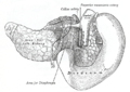
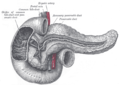
The second part, or descending part, of the duodenum begins at the superior duodenal flexure. It goes inferior to the lower border of vertebral body L3, before making a sharp turn medially into the inferior duodenal flexure, the end of the descending part.[8]: 274
The pancreatic duct and common bile duct enter the descending duodenum, through the major duodenal papilla. The second part of the duodenum also contains the minor duodenal papilla, the entrance for the accessory pancreatic duct. The junction between the embryological foregut and midgut lies just below the major duodenal papilla.[8]: 274
The third part, or horizontal part or inferior part of the duodenum is 10~12 cm in length. It begins at the inferior duodenal flexure and passes transversely to the left, passing in front of the inferior vena cava, abdominal aorta and the vertebral column. The superior mesenteric artery and vein are anterior to the third part of duodenum.[8]: 274 This part may be compressed between the aorta and SMA causing superior mesenteric artery syndrome.
The fourth part, or ascending part, of the duodenum passes upward, joining with the jejunum at the duodenojejunal flexure. The fourth part of the duodenum is at the vertebral level L3, and may pass directly on top, or slightly to the left, of the aorta.[8]: 274
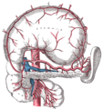
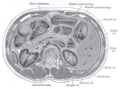
Blood supply[edit]
The duodenum receives arterial blood from two different sources. The transition between these sources is important as it demarcates the foregut from the midgut. Proximal to the 2nd part of the duodenum (approximately at the major duodenal papilla – where the bile duct enters) the arterial supply is from the gastroduodenal artery and its branch the superior pancreaticoduodenal artery. Distal to this point (the midgut) the arterial supply is from the superior mesenteric artery (SMA), and its branch the inferior pancreaticoduodenal artery supplies the 3rd and 4th sections.
The superior and inferior pancreaticoduodenal arteries (from the gastroduodenal artery and SMA respectively) form an anastomotic loop between the celiac trunk and the SMA; so there is potential for collateral circulation here.
The venous drainage of the duodenum follows the arteries. Ultimately these veins drain into the portal system, either directly or indirectly through the splenic or superior mesenteric vein and then to portal vein.
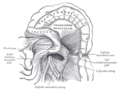
Lymphatic drainage[edit]
The lymphatic vessels follow the arteries in a retrograde fashion. The anterior lymphatic vessels drain into the pancreatoduodenal lymph nodes located along the superior and inferior pancreatoduodenal arteries and then into the pyloric lymph nodes (along the gastroduodenal artery). The posterior lymphatic vessels pass posterior to the head of the pancreas and drain into the superior mesenteric lymph nodes. Efferent lymphatic vessels from the duodenal lymph nodes ultimately pass into the celiac lymph nodes.
Histology[edit]
Under microscopy, the duodenum has a villous mucosa. This is distinct from the mucosa of the pylorus, which directly joins to the duodenum. Like other structures of the gastrointestinal tract, the duodenum has a mucosa, submucosa, muscularis externa, and adventitia. Glands line the duodenum, known as Brunner’s glands, which secrete mucus and bicarbonate in order to neutralise stomach acids. These are distinct glands not found in the ileum or jejunum, the other parts of the small intestine.[10]: 274–275
-

Dog Duodenum 100X
-
Duodenum with amyloid deposition in lamina propria
-

Section of duodenum of cat. X 60


Variation[edit]
|
This section needs expansion. You can help by adding to it. (December 2013) |
Gene and protein expression[edit]
About 20,000 protein coding genes are expressed in human cells and 70% of these genes are expressed in the normal duodenum.[11][12] Some 300 of these genes are more specifically expressed in the duodenum with very few genes expressed only in the duodenum. The corresponding specific proteins are expressed in the duodenal mucosa, and many of these are also expressed in the small intestine, such as alanine aminopeptidase, a digestive enzyme, angiotensin-converting enzyme, involved in controlling blood pressure, and RBP2, a protein involved in the uptake of vitamin A.[13]
Function[edit]
The duodenum is largely responsible for the breakdown of food in the small intestine, using enzymes. The duodenum also regulates the rate of emptying of the stomach via hormonal pathways. Secretin and cholecystokinin are released from cells in the duodenal epithelium in response to acidic and fatty stimuli present there when the pylorus opens and emits gastric chyme into the duodenum for further digestion. These cause the liver and gallbladder to release bile, and the pancreas to release bicarbonate and digestive enzymes such as trypsin, lipase and amylase into the duodenum as they are needed.
The villi of the duodenum have a leafy-looking appearance, which is a histologically identifiable structure. Brunner’s glands, which secrete mucus, are found in the duodenum only. The duodenum wall consists of a very thin layer of cells that form the muscularis mucosae.
Clinical significance[edit]
Ulceration[edit]
Ulcers of the duodenum commonly occur because of infection by the bacteria Helicobacter pylori. These bacteria, through a number of mechanisms, erode the protective mucosa of the duodenum, predisposing it to damage from gastric acids. The first part of the duodenum is the most common location of ulcers since it is where the acidic chyme meets the duodenal mucosa before mixing with the alkaline secretions of the duodenum.[14] Duodenal ulcers may cause recurrent abdominal pain and dyspepsia, and are often investigated using a urea breath test to test for the bacteria, and endoscopy to confirm ulceration and take a biopsy. If managed, these are often managed through antibiotics that aim to eradicate the bacteria, and proton-pump inhibitors and antacids to reduce the gastric acidity.[15]
Celiac disease[edit]
The British Society of Gastroenterology guidelines specify that a duodenal biopsy is required for the diagnosis of adult celiac disease. The biopsy is ideally performed at a moment when the patient is on a gluten-containing diet.[16]
Cancer[edit]
Duodenal cancer is a cancer in the first section of the small intestine. Cancer of the duodenum is relatively rare compared to stomach cancer and colorectal cancer; malignant tumors in the duodenum constitute only around 0.3% of all the gastrointestinal tract tumors but around half of cancerous tissues that develop in the small intestine.[17] Its histology is often observed to be adenocarcinoma, meaning that the cancerous tissue arises from glandular cells in the epithelial tissue lining the duodenum.[18]
Inflammation[edit]
Inflammation of the duodenum is referred to as duodenitis. There are multiple known causes.[19]
History[edit]
The name duodenum is from Medieval Latin, short for intestīnum duodēnum digitōrum, which may be translated: intestine of twelve finger-widths (in length), from Latin duodēnum, genitive pl. of duodēnī, twelve each, from duodecim, twelve.[20] The Latin phrase intestīnum duodēnum digitōrum is thought to be a loan-translation from the Greek word dodekadaktylon (δωδεκαδάκτυλον), literally «twelve fingers long.» The intestinal section was so called by Greek physician Herophilus (c. 335–280 BCE) for its length, about equal to the breadth of 12 fingers.[21]
Many languages retain a similar etymology for this word. For example, German Zwölffingerdarm and Dutch Twaalfvingerige darm.
Additional images[edit]
-

Sections of the small intestine
-

The celiac artery and its branches; the stomach has been raised and the peritoneum removed
-

Superior and inferior duodenal fossæ
-

Duodenojejunal fossa
-

The pancreas and duodenum from behind
-
Transverse section through the middle of the first lumbar vertebra, showing the relations of the pancreas
-
The pancreatic duct
-

Region of pancreas
-
Duodenum
-
Duodenum
-
Duodenum


See also[edit]
![]()
Wikimedia Commons has media related to Duodenum.
![]()
Look up duodenum in Wiktionary, the free dictionary.
- Pancreas
- Choledochoduodenostomy — a surgical procedure to create a connection between the common bile duct (CBD) and an alternative portion of the duodenum.
References[edit]
- ^ Nosek, Thomas M. «Section 6/6ch2/s6ch2_30». Essentials of Human Physiology. Archived from the original on 2016-03-24.
- ^ «NCI Dictionary of Cancer Terms». National Cancer Institute. Retrieved 2022-06-07.
The first part of the small intestine. It connects to the stomach. The duodenum helps to further digest food coming from the stomach. It absorbs nutrients (vitamins, minerals, carbohydrates, fats, proteins) and water from food so they can be used by the body.
- ^ «Duodenum: MedlinePlus Medical Encyclopedia». MedlinePlus. Retrieved 2022-06-07.
It is located between the stomach and the middle part of the small intestine. After foods mix with stomach acid, they move into the duodenum, where they mix with bile from the gallbladder and digestive juices from the pancreas.
- ^ a b Nolan, D. J. (2002). «Radiology of the Duodenum». Radiological Imaging of the Small Intestine. Medical Radiology. Berlin, Heidelberg: Springer Berlin Heidelberg. pp. 247–259. doi:10.1007/978-3-642-56231-0_6. ISBN 978-3-642-62993-8. ISSN 0942-5373.
duodenum is a C-shaped hollow organ forming an incomplete circle around the head of the pancreas. …it is normally examined as part of the upper gastrointestinal tract.
- ^ van Gijn J; Gijselhart JP (2011). «Treitz and his ligament». Ned. Tijdschr. Geneeskd. 155 (8): A2879. PMID 21557825.
- ^
Guillaume, Jean; Praxis Publishing; Sadasivam Kaushik; Pierre Bergot; Robert Metailler (2001). Nutrition and Feeding of Fish and Crustaceans. Springer. p. 31. ISBN 978-1-85233-241-9. Retrieved 2009-01-09. - ^ Latunde-Dada GO; Van der Westhuizen J; Vulpe CD; et al. (2002). «Molecular and functional roles of duodenal cytochrome B (Dcytb) in iron metabolism». Blood Cells Mol. Dis. 29 (3): 356–60. doi:10.1006/bcmd.2002.0574. PMID 12547225.
- ^ a b c d e f Drake, Richard L.; Vogl, Wayne; Tibbitts, Adam W.M. Mitchell; illustrations by Richard; Richardson, Paul (2005). Gray’s anatomy for students. Philadelphia: Elsevier/Churchill Livingstone. ISBN 978-0-8089-2306-0.
- ^ Singh, Inderbir; GP Pal (2012). «13». Human Embryology (9 ed.). Delhi: Macmillan Publishers India. p. 163. ISBN 978-93-5059-122-2.
- ^ Deakin, Barbara Young; et al. (2006). Wheater’s functional histology : a text and colour atlas (5th ed.). [Edinburgh?]: Churchill Livingstone/Elsevier. ISBN 978-0-443-06850-8.
- ^ «The human proteome in duodenum — The Human Protein Atlas». www.proteinatlas.org. Retrieved 2017-09-26.
- ^ Uhlén, Mathias; Fagerberg, Linn; Hallström, Björn M.; Lindskog, Cecilia; Oksvold, Per; Mardinoglu, Adil; Sivertsson, Åsa; Kampf, Caroline; Sjöstedt, Evelina (2015-01-23). «Tissue-based map of the human proteome». Science. 347 (6220): 1260419. doi:10.1126/science.1260419. ISSN 0036-8075. PMID 25613900. S2CID 802377.
- ^ Gremel, Gabriela; Wanders, Alkwin; Cedernaes, Jonathan; Fagerberg, Linn; Hallström, Björn; Edlund, Karolina; Sjöstedt, Evelina; Uhlén, Mathias; Pontén, Fredrik (2015-01-01). «The human gastrointestinal tract-specific transcriptome and proteome as defined by RNA sequencing and antibody-based profiling». Journal of Gastroenterology. 50 (1): 46–57. doi:10.1007/s00535-014-0958-7. ISSN 0944-1174. PMID 24789573. S2CID 21302849.
- ^ Smith, Margaret E. The Digestive System.
- ^ Britton, the editors Nicki R. Colledge, Brian R. Walker, Stuart H. Ralston; illustrated by Robert (2010). Davidson’s principles and practice of medicine (21st ed.). Edinburgh: Churchill Livingstone/Elsevier. pp. 871–874. ISBN 978-0-7020-3085-7.
- ^ Ludvigsson, J. F.; Bai, J. C.; Biagi, F.; Card, T. R.; Ciacci, C.; Ciclitira, P. J.; Green, P. H. R.; Hadjivassiliou, M.; Holdoway, A.; van Heel, D. A.; Kaukinen, K.; Leffler, D. A.; Leonard, J. N.; Lundin, K. E. A.; McGough, N.; Davidson, M.; Murray, J. A.; Swift, G. L.; Walker, M. M.; Zingone, F.; Sanders, D. S. (2014). «Diagnosis and management of adult coeliac disease: Guidelines from the British Society of Gastroenterology». Gut. 63 (8): 1210–1228. doi:10.1136/gutjnl-2013-306578. ISSN 0017-5749. PMC 4112432. PMID 24917550.
- ^ Fagniez, Pierre-Louis; Rotman, Nelly (2001). Malignant tumors of the duodenum. Zuckschwerdt.
- ^ https://www.cancer.gov/publications/dictionaries/cancer-terms/def/adenocarcinoma.
- ^ Serra S, Jani PA (2006). «An approach to duodenal biopsies». J. Clin. Pathol. 59 (11): 1133–50. doi:10.1136/jcp.2005.031260. PMC 1860495. PMID 16679353.
- ^ American Heritage Dictionary, 4th edition
- ^ «duodenum — Origin and meaning of duodenum by Online Etymology Dictionary». www.etymonline.com.
External links[edit]
![]()
Look up duodenum in Wiktionary, the free dictionary.
- Duodenum at the Human Protein Atlas
- duodenum at The Anatomy Lesson by Wesley Norman (Georgetown University)
From Wikipedia, the free encyclopedia
| Duodenum | |
|---|---|
|
Image of the gastrointestinal tract, with the duodenum highlighted. |
|
|
Diagram of the human duodenum with major parts labelled |
|
| Details | |
| Pronunciation | |
| Precursor | Foregut (1st and 2nd parts), Midgut (3rd and 4th part) |
| Part of | Small intestine |
| System | Digestive system |
| Artery | Inferior pancreaticoduodenal artery, Superior pancreaticoduodenal artery |
| Vein | Pancreaticoduodenal veins |
| Nerve | celiac ganglia, vagus[1] |
| Identifiers | |
| Latin | Intestinum duodenum |
| MeSH | D004386 |
| TA98 | A05.6.02.001 |
| TA2 | 2944 |
| FMA | 7206 |
| Anatomical terminology
[edit on Wikidata] |
The duodenum is the first section of the small intestine[2] in most higher vertebrates, including mammals, reptiles, and birds. In mammals it may be the principal site for iron absorption.
The duodenum precedes the jejunum and ileum and is the shortest part of the small intestine.
In humans, the duodenum is a hollow jointed tube about 25–38 centimetres (10–15 inches) long connecting the stomach to the middle part of the small intestine.[3][4] It begins with the duodenal bulb and ends at the suspensory muscle of duodenum.[5] Duodenum can be divided into four parts: the first (superior), the second (descending), the third (horizontal) and the fourth (ascending) parts.[4]
Overview[edit]
The duodenum is the first section of the small intestine in most higher vertebrates, including mammals, reptiles, and birds. In fish, the divisions of the small intestine are not as clear, and the terms anterior intestine or proximal intestine may be used instead of duodenum.[6] In mammals the duodenum may be the principal site for iron absorption.[7]
In humans, the duodenum is a C-shaped hollow jointed tube, 25–38 centimetres (10–15 inches) in length, lying adjacent to the stomach (and connecting it to the small intestine). It is divided anatomically into four sections. The first part lies within the peritoneum but its other parts are retroperitoneal.[8]: 273
Parts[edit]
The first part, or superior part, of the duodenum is a continuation from the pylorus to transpyloric plane. It is superior to the rest of the segments, at the vertebral level of L1. The duodenal bulb, about 2 cm (3⁄4 in) long, is the first part of the duodenum and is slightly dilated. The duodenal bulb is a remnant of the mesoduodenum, a mesentery that suspends the organ from the posterior abdominal wall in fetal life.[9] The first part of the duodenum is mobile, and connected to the liver by the hepatoduodenal ligament of the lesser omentum. The first part of the duodenum ends at the corner, the superior duodenal flexure.[8]: 273
Relations:[citation needed]
- Anterior
- Gallbladder
- Quadrate lobe of liver
- Posterior
- Bile duct
- Gastroduodenal artery
- Portal vein
- Inferior vena cava
- Head of pancreas
- Superior
- Neck of gallbladder
- Hepatoduodenal ligament (lesser omentum)
- Inferior
- Neck of pancreas
- Greater omentum
- Head of pancreas
The second part, or descending part, of the duodenum begins at the superior duodenal flexure. It goes inferior to the lower border of vertebral body L3, before making a sharp turn medially into the inferior duodenal flexure, the end of the descending part.[8]: 274
The pancreatic duct and common bile duct enter the descending duodenum, through the major duodenal papilla. The second part of the duodenum also contains the minor duodenal papilla, the entrance for the accessory pancreatic duct. The junction between the embryological foregut and midgut lies just below the major duodenal papilla.[8]: 274
The third part, or horizontal part or inferior part of the duodenum is 10~12 cm in length. It begins at the inferior duodenal flexure and passes transversely to the left, passing in front of the inferior vena cava, abdominal aorta and the vertebral column. The superior mesenteric artery and vein are anterior to the third part of duodenum.[8]: 274 This part may be compressed between the aorta and SMA causing superior mesenteric artery syndrome.
The fourth part, or ascending part, of the duodenum passes upward, joining with the jejunum at the duodenojejunal flexure. The fourth part of the duodenum is at the vertebral level L3, and may pass directly on top, or slightly to the left, of the aorta.[8]: 274
Blood supply[edit]
The duodenum receives arterial blood from two different sources. The transition between these sources is important as it demarcates the foregut from the midgut. Proximal to the 2nd part of the duodenum (approximately at the major duodenal papilla – where the bile duct enters) the arterial supply is from the gastroduodenal artery and its branch the superior pancreaticoduodenal artery. Distal to this point (the midgut) the arterial supply is from the superior mesenteric artery (SMA), and its branch the inferior pancreaticoduodenal artery supplies the 3rd and 4th sections.
The superior and inferior pancreaticoduodenal arteries (from the gastroduodenal artery and SMA respectively) form an anastomotic loop between the celiac trunk and the SMA; so there is potential for collateral circulation here.
The venous drainage of the duodenum follows the arteries. Ultimately these veins drain into the portal system, either directly or indirectly through the splenic or superior mesenteric vein and then to portal vein.
Lymphatic drainage[edit]
The lymphatic vessels follow the arteries in a retrograde fashion. The anterior lymphatic vessels drain into the pancreatoduodenal lymph nodes located along the superior and inferior pancreatoduodenal arteries and then into the pyloric lymph nodes (along the gastroduodenal artery). The posterior lymphatic vessels pass posterior to the head of the pancreas and drain into the superior mesenteric lymph nodes. Efferent lymphatic vessels from the duodenal lymph nodes ultimately pass into the celiac lymph nodes.
Histology[edit]
Under microscopy, the duodenum has a villous mucosa. This is distinct from the mucosa of the pylorus, which directly joins to the duodenum. Like other structures of the gastrointestinal tract, the duodenum has a mucosa, submucosa, muscularis externa, and adventitia. Glands line the duodenum, known as Brunner’s glands, which secrete mucus and bicarbonate in order to neutralise stomach acids. These are distinct glands not found in the ileum or jejunum, the other parts of the small intestine.[10]: 274–275
-
Dog Duodenum 100X
-
Duodenum with amyloid deposition in lamina propria
-
Section of duodenum of cat. X 60
Variation[edit]
|
This section needs expansion. You can help by adding to it. (December 2013) |
Gene and protein expression[edit]
About 20,000 protein coding genes are expressed in human cells and 70% of these genes are expressed in the normal duodenum.[11][12] Some 300 of these genes are more specifically expressed in the duodenum with very few genes expressed only in the duodenum. The corresponding specific proteins are expressed in the duodenal mucosa, and many of these are also expressed in the small intestine, such as alanine aminopeptidase, a digestive enzyme, angiotensin-converting enzyme, involved in controlling blood pressure, and RBP2, a protein involved in the uptake of vitamin A.[13]
Function[edit]
The duodenum is largely responsible for the breakdown of food in the small intestine, using enzymes. The duodenum also regulates the rate of emptying of the stomach via hormonal pathways. Secretin and cholecystokinin are released from cells in the duodenal epithelium in response to acidic and fatty stimuli present there when the pylorus opens and emits gastric chyme into the duodenum for further digestion. These cause the liver and gallbladder to release bile, and the pancreas to release bicarbonate and digestive enzymes such as trypsin, lipase and amylase into the duodenum as they are needed.
The villi of the duodenum have a leafy-looking appearance, which is a histologically identifiable structure. Brunner’s glands, which secrete mucus, are found in the duodenum only. The duodenum wall consists of a very thin layer of cells that form the muscularis mucosae.
Clinical significance[edit]
Ulceration[edit]
Ulcers of the duodenum commonly occur because of infection by the bacteria Helicobacter pylori. These bacteria, through a number of mechanisms, erode the protective mucosa of the duodenum, predisposing it to damage from gastric acids. The first part of the duodenum is the most common location of ulcers since it is where the acidic chyme meets the duodenal mucosa before mixing with the alkaline secretions of the duodenum.[14] Duodenal ulcers may cause recurrent abdominal pain and dyspepsia, and are often investigated using a urea breath test to test for the bacteria, and endoscopy to confirm ulceration and take a biopsy. If managed, these are often managed through antibiotics that aim to eradicate the bacteria, and proton-pump inhibitors and antacids to reduce the gastric acidity.[15]
Celiac disease[edit]
The British Society of Gastroenterology guidelines specify that a duodenal biopsy is required for the diagnosis of adult celiac disease. The biopsy is ideally performed at a moment when the patient is on a gluten-containing diet.[16]
Cancer[edit]
Duodenal cancer is a cancer in the first section of the small intestine. Cancer of the duodenum is relatively rare compared to stomach cancer and colorectal cancer; malignant tumors in the duodenum constitute only around 0.3% of all the gastrointestinal tract tumors but around half of cancerous tissues that develop in the small intestine.[17] Its histology is often observed to be adenocarcinoma, meaning that the cancerous tissue arises from glandular cells in the epithelial tissue lining the duodenum.[18]
Inflammation[edit]
Inflammation of the duodenum is referred to as duodenitis. There are multiple known causes.[19]
History[edit]
The name duodenum is from Medieval Latin, short for intestīnum duodēnum digitōrum, which may be translated: intestine of twelve finger-widths (in length), from Latin duodēnum, genitive pl. of duodēnī, twelve each, from duodecim, twelve.[20] The Latin phrase intestīnum duodēnum digitōrum is thought to be a loan-translation from the Greek word dodekadaktylon (δωδεκαδάκτυλον), literally «twelve fingers long.» The intestinal section was so called by Greek physician Herophilus (c. 335–280 BCE) for its length, about equal to the breadth of 12 fingers.[21]
Many languages retain a similar etymology for this word. For example, German Zwölffingerdarm and Dutch Twaalfvingerige darm.
Additional images[edit]
-
Sections of the small intestine
-
The celiac artery and its branches; the stomach has been raised and the peritoneum removed
-
Superior and inferior duodenal fossæ
-
Duodenojejunal fossa
-
The pancreas and duodenum from behind
-
Transverse section through the middle of the first lumbar vertebra, showing the relations of the pancreas
-
The pancreatic duct
-
Region of pancreas
-
Duodenum
-
Duodenum
-
Duodenum
See also[edit]
![]()
Wikimedia Commons has media related to Duodenum.
![]()
Look up duodenum in Wiktionary, the free dictionary.
- Pancreas
- Choledochoduodenostomy — a surgical procedure to create a connection between the common bile duct (CBD) and an alternative portion of the duodenum.
References[edit]
- ^ Nosek, Thomas M. «Section 6/6ch2/s6ch2_30». Essentials of Human Physiology. Archived from the original on 2016-03-24.
- ^ «NCI Dictionary of Cancer Terms». National Cancer Institute. Retrieved 2022-06-07.
The first part of the small intestine. It connects to the stomach. The duodenum helps to further digest food coming from the stomach. It absorbs nutrients (vitamins, minerals, carbohydrates, fats, proteins) and water from food so they can be used by the body.
- ^ «Duodenum: MedlinePlus Medical Encyclopedia». MedlinePlus. Retrieved 2022-06-07.
It is located between the stomach and the middle part of the small intestine. After foods mix with stomach acid, they move into the duodenum, where they mix with bile from the gallbladder and digestive juices from the pancreas.
- ^ a b Nolan, D. J. (2002). «Radiology of the Duodenum». Radiological Imaging of the Small Intestine. Medical Radiology. Berlin, Heidelberg: Springer Berlin Heidelberg. pp. 247–259. doi:10.1007/978-3-642-56231-0_6. ISBN 978-3-642-62993-8. ISSN 0942-5373.
duodenum is a C-shaped hollow organ forming an incomplete circle around the head of the pancreas. …it is normally examined as part of the upper gastrointestinal tract.
- ^ van Gijn J; Gijselhart JP (2011). «Treitz and his ligament». Ned. Tijdschr. Geneeskd. 155 (8): A2879. PMID 21557825.
- ^
Guillaume, Jean; Praxis Publishing; Sadasivam Kaushik; Pierre Bergot; Robert Metailler (2001). Nutrition and Feeding of Fish and Crustaceans. Springer. p. 31. ISBN 978-1-85233-241-9. Retrieved 2009-01-09. - ^ Latunde-Dada GO; Van der Westhuizen J; Vulpe CD; et al. (2002). «Molecular and functional roles of duodenal cytochrome B (Dcytb) in iron metabolism». Blood Cells Mol. Dis. 29 (3): 356–60. doi:10.1006/bcmd.2002.0574. PMID 12547225.
- ^ a b c d e f Drake, Richard L.; Vogl, Wayne; Tibbitts, Adam W.M. Mitchell; illustrations by Richard; Richardson, Paul (2005). Gray’s anatomy for students. Philadelphia: Elsevier/Churchill Livingstone. ISBN 978-0-8089-2306-0.
- ^ Singh, Inderbir; GP Pal (2012). «13». Human Embryology (9 ed.). Delhi: Macmillan Publishers India. p. 163. ISBN 978-93-5059-122-2.
- ^ Deakin, Barbara Young; et al. (2006). Wheater’s functional histology : a text and colour atlas (5th ed.). [Edinburgh?]: Churchill Livingstone/Elsevier. ISBN 978-0-443-06850-8.
- ^ «The human proteome in duodenum — The Human Protein Atlas». www.proteinatlas.org. Retrieved 2017-09-26.
- ^ Uhlén, Mathias; Fagerberg, Linn; Hallström, Björn M.; Lindskog, Cecilia; Oksvold, Per; Mardinoglu, Adil; Sivertsson, Åsa; Kampf, Caroline; Sjöstedt, Evelina (2015-01-23). «Tissue-based map of the human proteome». Science. 347 (6220): 1260419. doi:10.1126/science.1260419. ISSN 0036-8075. PMID 25613900. S2CID 802377.
- ^ Gremel, Gabriela; Wanders, Alkwin; Cedernaes, Jonathan; Fagerberg, Linn; Hallström, Björn; Edlund, Karolina; Sjöstedt, Evelina; Uhlén, Mathias; Pontén, Fredrik (2015-01-01). «The human gastrointestinal tract-specific transcriptome and proteome as defined by RNA sequencing and antibody-based profiling». Journal of Gastroenterology. 50 (1): 46–57. doi:10.1007/s00535-014-0958-7. ISSN 0944-1174. PMID 24789573. S2CID 21302849.
- ^ Smith, Margaret E. The Digestive System.
- ^ Britton, the editors Nicki R. Colledge, Brian R. Walker, Stuart H. Ralston; illustrated by Robert (2010). Davidson’s principles and practice of medicine (21st ed.). Edinburgh: Churchill Livingstone/Elsevier. pp. 871–874. ISBN 978-0-7020-3085-7.
- ^ Ludvigsson, J. F.; Bai, J. C.; Biagi, F.; Card, T. R.; Ciacci, C.; Ciclitira, P. J.; Green, P. H. R.; Hadjivassiliou, M.; Holdoway, A.; van Heel, D. A.; Kaukinen, K.; Leffler, D. A.; Leonard, J. N.; Lundin, K. E. A.; McGough, N.; Davidson, M.; Murray, J. A.; Swift, G. L.; Walker, M. M.; Zingone, F.; Sanders, D. S. (2014). «Diagnosis and management of adult coeliac disease: Guidelines from the British Society of Gastroenterology». Gut. 63 (8): 1210–1228. doi:10.1136/gutjnl-2013-306578. ISSN 0017-5749. PMC 4112432. PMID 24917550.
- ^ Fagniez, Pierre-Louis; Rotman, Nelly (2001). Malignant tumors of the duodenum. Zuckschwerdt.
- ^ https://www.cancer.gov/publications/dictionaries/cancer-terms/def/adenocarcinoma.
- ^ Serra S, Jani PA (2006). «An approach to duodenal biopsies». J. Clin. Pathol. 59 (11): 1133–50. doi:10.1136/jcp.2005.031260. PMC 1860495. PMID 16679353.
- ^ American Heritage Dictionary, 4th edition
- ^ «duodenum — Origin and meaning of duodenum by Online Etymology Dictionary». www.etymonline.com.
External links[edit]
![]()
Look up duodenum in Wiktionary, the free dictionary.
- Duodenum at the Human Protein Atlas
- duodenum at The Anatomy Lesson by Wesley Norman (Georgetown University)


Орфографический словарь русского языка (онлайн)
Как пишется слово «двенадцатиперстная кишка» ?
Правописание слова «двенадцатиперстная кишка»
А Б В Г Д Е Ж З И Й К Л М Н О П Р С Т У Ф Х Ц Ч Ш Щ Э Ю Я
двенадцатипе́рстная кишка́
Рядом по алфавиту:
ДВ-диапазо́н , -а
два́-три́ , дву́х-трёх
двенадцатери́чный
двенадцатиби́тный , (12-би́тный)
двенадцатигра́нник , -а
двенадцатигра́нный
двенадцатидне́вный , (12-дне́вный)
двенадцатидюймо́вка , -и, р. мн. -вок
двенадцатидюймо́вый , (12-дюймо́вый)
двенадцатиза́льный , (12-за́льный)
двенадцатизву́чный , (12-зву́чный)
двенадцатиле́тка , -и, р. мн. -ток
двенадцатиле́тний , (12-ле́тний)
двенадцатиме́сячный , (12-ме́сячный)
двенадцатими́льный , (12-ми́льный)
двенадцатипе́рстная кишка́
двенадцатипе́рстно-тощекише́чный
двенадцатирублёвый , (12-рублёвый)
двенадцатиря́дный
двенадцатисве́чник , -а
двенадцатисло́жный
двенадцатисти́шие , (12-сти́шие), -я
двенадцатито́новый , (12-то́новый)
двенадцатито́чечный жу́к
двенадцатиуго́льник , -а
двенадцатиуго́льный
двенадцатицили́ндровый , (12-цили́ндровый)
двенадцатичасово́й , (12-часово́й)
двенадцатиэта́жка , -и, р. мн. -жек
двенадцатиэта́жный , (12-эта́жный)
двена́дцатый
Содержание
- 1 Русский
- 1.1 Тип и синтаксические свойства сочетания
- 1.2 Произношение
- 1.3 Семантические свойства
- 1.3.1 Значение
- 1.3.2 Синонимы
- 1.3.3 Антонимы
- 1.3.4 Гиперонимы
- 1.3.5 Гипонимы
- 1.4 Этимология
- 1.5 Перевод
- 1.6 Библиография
Русский[править]
Тип и синтаксические свойства сочетания[править]
две—над—ца—ти—пе́р—стна·я киш—ка́
Устойчивое сочетание (термин). Используется в качестве именной группы.
Произношение[править]
- МФА: [dvʲɪˌnat͡s(ː)ətʲɪˈpʲersnəɪ̯ə kʲɪˈʂka]
Семантические свойства[править]

Значение[править]
- анат. часть тонкой кишки человека, начинающаяся от желудка ◆ Отсутствует пример употребления (см. рекомендации).
Синонимы[править]
Антонимы[править]
Гиперонимы[править]
Гипонимы[править]
Этимология[править]
Калька нем. Zwölffingerdarm, передающего лат. intestinum duodenum digitorum. Название дано по длине этой кишки, равной около 12 пальцев (перстов) в поперечнике.
Перевод[править]
| Список переводов | |
|
Библиография[править]
- Аникин А. Е. Русский этимологический словарь. — М. : Рукописные памятники Древней Руси, 2007—. — Т. 13.