![]()
Слова русского языка,
поиск и разбор слов онлайн
гипофиз
Правильно слово пишется: гипо́фи́з
В этом слове допускается ставить ударение на разные слоги.
Всего в слове 7 букв, 3 гласных, 4 согласных, 3 слога.
Гласные: и, о, и;
Согласные: г, п, ф, з.
Номера букв в слове
Номера букв в слове «гипофиз» в прямом и обратном порядке:
- 7
г
1 - 6
и
2 - 5
п
3 - 4
о
4 - 3
ф
5 - 2
и
6 - 1
з
7
Как правильно пишется слово «гипофиз»
гипо́фи́з
гипо́фи́з, -а
Источник: Орфографический
академический ресурс «Академос» Института русского языка им. В.В. Виноградова РАН (словарная база
2020)
Делаем Карту слов лучше вместе

Привет! Меня зовут Лампобот, я компьютерная программа, которая помогает делать
Карту слов. Я отлично
умею считать, но пока плохо понимаю, как устроен ваш мир. Помоги мне разобраться!
Спасибо! Я стал чуточку лучше понимать мир эмоций.
Вопрос: трувер — это что-то нейтральное, положительное или отрицательное?
Ассоциации к слову «гипофиз»
Синонимы к слову «гипофиз»
Предложения со словом «гипофиз»
- Тиреотропный гормон гипофиза управляет деятельностью щитовидной железы.
- В норме это приводит к снижению выработки тиреотропина, а значит, и работы гипофиза.
- Гипофиз начинает отслеживать внешние входящие сигналы и по ним работать.
- (все предложения)
Значение слова «гипофиз»
-
ГИПО́ФИЗ и ГИПОФИ́З, -а, м. Анат. Железа внутренней секреции, расположенная у основания головного мозга, оказывающая влияние на рост и развитие организма. (Малый академический словарь, МАС)
Все значения слова ГИПОФИЗ
Отправить комментарий
Дополнительно
Синонимы слова «ГИПОФИЗ»:
ЖЕЛЕЗА, ПИТУИТАРНАЯ ЖЕЛЕЗА, ПРИДАТОК
Смотреть что такое ГИПОФИЗ в других словарях:
ГИПОФИЗ
(от греч. hypóphysis — отросток) нижний мозговой придаток (hypophysis cerebri, glandula pituitaria), железа с внутренней секрецией (См. Внутренн… смотреть
ГИПОФИЗ
гипофиз м. Железа внутренней секреции, расположенная у основания головного мозга человека и позвоночных животных и оказывающая влияние на рост, развитие, обменные процессы и т.п. организма; мозговой придаток.<br><br><br>… смотреть
ГИПОФИЗ
гипофиз
железа, придаток, питуитарная железа
Словарь русских синонимов.
гипофиз
сущ., кол-во синонимов: 3
• железа (20)
• питуитарная железа (1)
• придаток (13)
Словарь синонимов ASIS.В.Н. Тришин.2013.
.
Синонимы:
железа, питуитарная железа, придаток… смотреть
ГИПОФИЗ
IГипо́физ (hypophysis, glandula pituitana. греч hypo- + phyō, будущее время physō расти; синоним: мозговой придаток, питуитарная железа)железа внутренн… смотреть
ГИПОФИЗ
ГИПОФИЗили нижний мозговой придаток, эндокринная железа, расположенная в костном кармане (турецком седле) у основания мозга. У человека он величиной с горошину и весит около 0,5 г.Гипофиз состоит из трех долей: передней, промежуточной и задней. Первые две доли состоят из железистой ткани и образуются у эмбриона из кармана Ратке — переднего выпячивания кишечной трубки. Заднюю долю образует вырост нервной ткани, идущий от дна промежуточного мозга. Все эти доли фактически являются отдельными железами, и каждая секретирует свои собственные гормоны.Передняя доля гипофиза вырабатывает белковые гормоны, шесть из которых выделены в химически чистом виде. Их строение в настоящее время полностью расшифровано. Точное число секретируемых передней долей гормонов не установлено, ниже рассматриваются лишь хорошо известные.Гормон роста. На рост организма влияют многие гормоны, но наиболее важную роль в этом сложном процессе играет, видимо, именно гипофизарный гормон роста (соматотропин). После удаления гипофиза рост практически прекращается. Введение этого гормона молодым животным ускоряет рост, а у взрослых может приводить к его возобновлению, причем исследование обмена веществ в этих случаях всегда выявляет снижение экскреции (выведения) азота из организма. Задержка азота — необходимый признак истинного роста, свидетельствующий о том, что действительно происходит образование новых тканей, а не просто увеличение массы тела за счет накопления жира или воды. При патологических процессах, ведущих к снижению функции гипофиза, в отдельных случаях возникает гипофизарная карликовость; такие карлики имеют небольшие размеры тела, но в остальном остаются нормальными людьми. Другие нарушения функции гипофиза могут сопровождаться избыточным выделением гормона роста, порождающим гигантизм. Если большие количества гормона роста вырабатываются до завершения созревания организма, рост увеличивается пропорционально; если же это происходит уже после достижения зрелости, возникает состояние, называемое акромегалией, при котором наблюдается непропорциональный рост отдельных частей тела, поскольку у взрослых некоторые кости теряют способность к дальнейшему удлинению. При акромегалии больной приобретает характерный внешний облик: начинают выдаваться брови, нос и нижняя челюсть, увеличиваются кисти рук, стопы и грудь, спина становится неподвижной, нос и губы утолщаются.Лактогенный гормон гипофиза (пролактин) стимулирует лактацию — образование молока в молочных железах. Стойкая лактация в сочетании с аменореей (аномальным отсутствием или подавлением менструальных выделений) может возникать при опухоли гипофиза. Это расстройство бывает также связано с нарушениями секреторной активности гипоталамуса, в норме подавляющей высвобождение пролактина. У самок некоторых млекопитающих пролактин влияет и на другие процессы, в частности он может стимулировать секрецию гормона прогестерона желтым телом яичника. Пролактин присутствует в гипофизе особей не только женского, но и мужского пола, причем не только у млекопитающих, но и у низших позвоночных. Относительно его функций в мужском организме и у животных, не относящихся к млекопитающим, известно мало. У некоторых птиц пролактин стимулирует развитие зобного мешка. Поскольку «зобное молоко», вырабатывающееся в этом мешке, служит для вскармливания птенцов, такое действие гормона функционально сходно с его действием у млекопитающих. У рыб пролактин участвует в регуляции осмотического давления крови.Тиреотропный гормон гипофиза (тиреотропин) стимулирует рост щитовидной железы и ее секреторную активность. После удаления гипофиза функция щитовидной железы полностью прекращается и она уменьшается в размерах. Введение тиреотропина может вызвать избыточную активность щитовидной железы. Таким образом, нарушения ее функции могут быть следствием не только заболеваний самой железы, но и патологических процессов в гипофизе и соответственно требуют разного лечения.Адренокортикотропный гормон гипофиза (АКТГ, кортикотропин) стимулирует кору надпочечников подобно тому, как тиреотропный гормон стимулирует щитовидную железу. Одно из различий, однако, заключается в том, что функция коры надпочечников в отсутствие АКТГ прекращается не полностью. Когда стимуляция со стороны гипофиза отсутствует, кора надпочечников сохраняет способность секретировать необходимый для жизни гормон альдостерон, который регулирует содержание натрия и калия в организме. Однако без АКТГ надпочечники вырабатывают недостаточное количество другого жизненно важного гормона, кортизола, и теряют способность усиливать при необходимости его секрецию. Поэтому больные с недостаточностью функции гипофиза становятся очень чувствительны к различного рода нагрузкам и стрессам. Избыточные количества АКТГ, которые могут вырабатываться при опухолях гипофиза, приводят к развитию потенциально смертельного заболевания, т.н. синдрома Кушинга. К характерным его признакам относятся прибавка в весе, лунообразное лицо, увеличение жировых отложений в верхней части туловища, повышение кровяного давления, мышечная слабость.Гонадотропные гормоны (гонадотропины). Передняя доля гипофиза секретирует два гонадотропных гормона. Один из них, фолликулостимулирующий гормон, стимулирует развитие яйцеклеток в яичниках и сперматозоидов в семенниках. Второй называется лютеинизирующим гормоном; в женском организме он стимулирует выработку в яичниках женских половых гормонов и выход зрелой яйцеклетки из яичника, а в мужском — секрецию гормона тестостерона интерстициальными клетками семенников (см. также РЕПРОДУКЦИЯ ЧЕЛОВЕКА). Введение этих гормонов или их избыточная продукция вследствие заболевания вызывают преждевременное половое развитие незрелого организма. При удалении гипофиза или его разрушении патологическим процессом возникают изменения, сходные с теми, что происходят при кастрации.Регуляция метаболизма. Гормоны, секретируемые передней долей гипофиза, необходимы для надлежащего использования в организме углеводов, поступающих с пищей; кроме того, они выполняют и другие важные функции в обмене веществ. Особая роль в регуляции метаболизма принадлежит, по-видимому, гормону роста и адренокортикотропному гормону, которые функционально тесно связаны с гормоном поджелудочной железы, инсулином. Хорошо известно, что в отсутствие инсулина развивается хроническое заболевание — сахарный диабет. При одновременном удалении поджелудочной железы и гипофиза большинство симптомов диабета отсутствует, так что в этом отношении влияние гормонов гипофиза и поджелудочной железы как бы противоположно.Промежуточная доля гипофиза секретирует меланоцит-стимулирующий гормон (МСГ, интермедин), который увеличивает размеры некоторых пигментных клеток в коже низших позвоночных. Например, лишенные этого гормона головастики из-за сокращения (сжатия) пигментных клеток приобретают серебристый цвет. МСГ образуется из той же молекулы-предшественника, что и адренокортикотропный гормон (АКТГ). В передней доле гипофиза этот предшественник превращается в АКТГ, а в промежуточной — в МСГ. МСГ вырабатывается и в гипофизе млекопитающих, но его функция остается неясной.Задняя доля гипофиза содержит два гормона, причем оба вырабатываются в гипоталамусе, а оттуда поступают в гипофиз. Один из них, окситоцин, — наиболее активный из присутствующих в организме факторов, вызывающий такие же сильные сокращения матки, как при родах. Этот гормон иногда применяют в акушерстве для стимуляции затянувшихся родов, но значение его нормальных концентраций в родовой деятельности не установлено. Окситоцин вызывает также сокращения мышечных стенок желчного пузыря, кишечника, мочеточников и мочевого пузыря. Второй гормон, вазопрессин, при введении в организм вызывает многочисленные эффекты, в том числе повышение кровяного давления вследствие сужения сосудов и уменьшение диуреза (выведения мочи). Однако в нормальных условиях он оказывает в организме лишь одно известное действие — регулирует количество воды, выделяющееся через почки. Даже под влиянием чрезвычайно малых его концентраций вода, профильтровавшаяся в почечных клубочках, всасывается обратно в почечных канальцах (реабсорбируется), и образуется концентрированная моча. При разрушении задней доли гипофиза опухолями или другими патологическими процессами развивается состояние, называемое несахарным диабетом. При этом заболевании организм теряет через почки огромное количество воды, превышающее иногда 38 л в сутки. Возникает сильная жажда, и чтобы избежать обезвоживания, больным приходится потреблять соответствующее количество воды. См. также ГОРМОНЫ; ЭНДОКРИННАЯ СИСТЕМА…. смотреть
ГИПОФИЗ
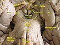
Микропрепарат аденогипофиза крупного рогатого скота.
Микропрепарат аденогипофиза крупного рогатого скота:1 базофильные клетки;2 ацидофильные клетк… смотреть
ГИПОФИЗ
(гип… +греч. phyo, будущее время physo — расти, синоним — мозговой придаток), железа внутренней секреции, расположенная в основании черепа и … смотреть
ГИПОФИЗ
ГИПОФИЗ
(от греч. hypophysis — отросток), нижний мозговой придаток, питуитарная железa (hypophysis cerebri, glandula pituitaria), железа внутр. секрец… смотреть
ГИПОФИЗ
Гипофиз (hypophysis) (рис. 244) представляет собой непарный орган округлой формы и залегает в гипофизарной ямке турецкого седла. Вес его составляет 0,5… смотреть
ГИПОФИЗ
гипо́физ
((гр. hypophysis отросток) нижний мозговой придаток, железа внутренней секреции с многообразными функциями; играет ведущую роль в гормонально… смотреть
ГИПОФИЗ
ГИПОФИЗ (лат. Hypophysis cerebri) — одна из желез внутренней секреции (т. е. эндокринная железа), прикрепленная к мозгу. Г. представляет собой непарный железисто-нервный орган (масса от 0,3 до 0,7 г), расположенный у основания мозга, под перекрестом оптических нервов (хиазмой), и спускающийся в углубление турецкого седла основной кости черепа. Г. часто называют нижним придатком мозга в противоположность эпифизу (шишковидное тело) — верхнему придатку мозга. В Г. различают 3 доли: переднюю — железистую (секретирующую 7 гормонов), среднюю (у человека недоразвита) и заднюю (секретирует вазопрессин и окситоцин, синтезируемые в гипоталамусе). Лат. названия: Glandula pituitaria, Colatorium, Labrum, Embotum, Sentina и др. Обилие названий отражает тот факт, что этому органу приписывали самые разные функции (рост, развитие, обмен веществ и др.). Действительно, в эндокринной системе Г. занимает особое положение, поскольку посредством своих гормонов оказывает регулирующие воздействия на др. железы и органы. Но и сам Г. находится под контролем гипоталамуса, который, в свою очередь, входит в систему «эмоционального мозга». Т. о., посредством дуэта гипоталамус—Г. эмоциональная жизнь человека может иметь сильное влияние на организм. В ответ на стрессовую ситуацию Г. выделает адренокортикотропный гормон (АКТГ), который влияет на секрецию гормонов коры надпочечников (см. Кортикостероиды). Г. также отвечает за синтез гормона роста (соматотропный гормон), гонадотропные гормоны, тиреотропный гормон. Нарушение функций Г. приводит к многим заболеваниям (напр., акромегалия, гигантизм, нанизм). (Б. М.)<br><br><br>… смотреть
ГИПОФИЗ
(лат. Hypophysis cerebri) — одна из желез внутренней секреции (т. е. эндокринная железа), прикрепленная к мозгу. Г. представляет собой непарный железисто-нервный орган (масса от 0,3 до 0,7 г), расположенный у основания мозга, под перекрестом оптических нервов (хиазмой), и спускающийся в углубление турецкого седла основной кости черепа. Г. часто называют нижним придатком мозга в противоположность эпифизу (шишковидное тело) — верхнему придатку мозга.В Г. различают 3 доли: переднюю — железистую (секретирующую 7 гормонов), среднюю (у человека недоразвита) и заднюю (секретирует вазопрессин и окситоцин, синтезируемые в гипоталамусе). Лат. названия: Glandula pituitaria, Colatorium, Labrum, Embotum, Sentina и др. Обилие названий отражает тот факт, что этому органу приписывали самые разные функции (рост, развитие, обмен веществ и др.). Действительно, в эндокринной системе Г. занимает особое положение, поскольку посредством своих гормонов оказывает регулирующие воздействия на др. железы и органы. Но и сам Г. находится под контролем гипоталамуса, который, в свою очередь, входит в систему «эмоционального мозга». Т. о., посредством дуэта гипоталамус—Г. эмоциональная жизнь человека может иметь сильное влияние на организм. В ответ на стрессовую ситуацию Г. выделает адренокортикотропный гормон (АКТГ), который влияет на секрецию гормонов коры надпочечников (см. Кортикостероиды). Г. также отвечает за синтез гормона роста (соматотропный гормон), гонадотропные гормоны, тиреотропный гормон. Нарушение функций Г. приводит к многим заболеваниям (напр., акромегалия, гигантизм, нанизм). (Б. М.)… смотреть
ГИПОФИЗ
расположенный под таламусом, то есть под зрительным бугром (греч. hypophysis отросток) “главная” эндокринная железа, контролирующая активность других эндокринных желёз. Установлено, что передняя доля гипофиза продуцирует: 1. соматотропный гормон (СТГ, гормон роста); 2. адренокортикотропный гормон (АКТГ, который регулирует деятельность надпочечников); 3. тиреотропный гормон (ТГ, регулирущий деятельность щитовидной железы); 4. гонадотропные гормоны (ГГ), такие, как: а) фолликулостимулирующий гормон (ФСГ, стимулирующий развитие фолликулов у женщин и сперматогенез у мужчин), б) лютеинизирующий гормон (ЛГ, который совместно с ФСГ стимулирует секрецию эстрогенов, овуляцию и развитие жёлтого тела) и в) лактогенный гормон (ЛГ, контролирующий продуцирование молока в зрелой молочной железе матери). В свою очередь, задняя доля гипофиза (нейрогипофиз) продуцирует: 5. антидиуретический гормон (АДГ, контролирует водный обмен), 6. вазопрессин (стимулирует сокращение гладкой мускулатуры кровеносных сосудов) и 7. окситоцин (усиливает сокращение матки и выделение молока молочными железами)…. смотреть
ГИПОФИЗ
(pityitary gland, hypophysis) одна из основных эндокринных желез в организме человека; напоминающее горошину образование, расположенное в гипофизарной ямке турецкого седла клиновидной кости. Воронка, проходящая через диафрагму седла, соединяет гипофиз с гипоталамусом. Клетки передней доли (аденогипофиза (adenohypophysis)) синтезируют тиреотропный гормон, АКТГ (адренокортикотропный гормон), гонадотропные гормоны (фолликулостимулирующий ФСГ и лютеинизирующий ЛГ (ред.)), гормон роста, пролактин, липотрофин и меланоцитостимулирующий гормон. Секреция всех этих гормонов регулируется специфическими освобождающими гормонами (releasing hormones) (рилизинг факторами, или либеринами, и ингибирующими гормонами, или статинами), которые образуются в гипоталамусе (см. также Гормон гонадотропин-освобождающий). Из задней доли гипофиза (нейрогипофиза (neurohypophysis)) в кровь выделяются вазопрессин и окситоцин, которые синтезируются в гипоталамусе и транспортируются в гипофиз, где хранятся до момента их освобождения…. смотреть
ГИПОФИЗ
ГИПОФИЗ, hypophysis, is, f (гр. hypo + phyo образование, возникновение) — эндокринная железа, расположенная у основания головного мозга, в турецком седле. Г. состоит из двух главных долей: из аденогипофиза (который возникает из стомадеальной впадины зародыша), и из нейрогипофиза (возникшего в виде выпячивания из дна промежуточного мозга). В аденогипофизе, в свою очередь, различают: инфундибулярную часть, охватывающую в области гипофизарной ножки проксимальную часть нейрогипофиза снаружи; промежуточную часть, между дистальными частями адено- и нейрогипофиза, и дистальную часть аденогипофиза. Нейрогипофиз состоит из супраселлярной проксимальной части и из дистальной части, занимающей место внутри седла. Аденогипофиз продуцирует несколько гормонов, которые регулируют правильное функционирование других эндокринных желез.<br><br><br>… смотреть
ГИПОФИЗ
железа внутр. секреции позвоночных животных и человека. Масса 0,5-0,6 г. Г. расположен у основания головного мозга и состоит из 2 долей: передней (аден… смотреть
ГИПОФИЗ
ГИПОФИЗ, железа внутренней секреции позвоночных животных и человека. Весит 0, 5-0, 6 г. Гипофиз расположен у основания головного мозга и состоит из 2 долей: передней (аденогипофиз) и задней (нейрогипофиз). Тесно связан с гипоталамусом, клетки которого вырабатывают рилизинг-гормоны, стимулирующие или угнетающие секрецию гормонов передней долей гипофиза (адренокортикотропного, лютеинизирующего, пролактина, соматотропного, фолликулостимулирующего и др.). Гормоны окситоцин и вазопрессин, выделяемые задней долей гипофиза, также образуются в гипоталамусе. Гипофиз оказывает преимущественное влияние на рост, развитие, обменные процессы, регулирует деятельность других желез внутренней секреции. Поражения гипофиза приводят к различным заболеваниям (напр., акромегалии, гигантизму).<br><br><br>… смотреть
ГИПОФИЗ
ГИПОФИЗ — железа внутренней секреции позвоночных животных и человека. Весит 0,5-0,6 г. Гипофиз расположен у основания головного мозга и состоит из 2 долей: передней (аденогипофиз) и задней (нейрогипофиз). Тесно связан с гипоталамусом, клетки которого вырабатывают рилизинг-гормоны, стимулирующие или угнетающие секрецию гормонов передней долей гипофиза (адренокортикотропного, лютеинизирующего, пролактина, соматотропного, фолликулостимулирующего и др.). Гормоны окситоцин и вазопрессин, выделяемые задней долей гипофиза, также образуются в гипоталамусе. Гипофиз оказывает преимущественное влияние на рост, развитие, обменные процессы, регулирует деятельность других желез внутренней секреции. Поражения гипофиза приводят к различным заболеваниям (напр., акромегалии, гигантизму).<br>… смотреть
ГИПОФИЗ
ГИПОФИЗ, железа внутренней секреции позвоночных животных и человека. Весит 0,5-0,6 г. Гипофиз расположен у основания головного мозга и состоит из 2 долей: передней (аденогипофиз) и задней (нейрогипофиз). Тесно связан с гипоталамусом, клетки которого вырабатывают рилизинг-гормоны, стимулирующие или угнетающие секрецию гормонов передней долей гипофиза (адренокортикотропного, лютеинизирующего, пролактина, соматотропного, фолликулостимулирующего и др.). Гормоны окситоцин и вазопрессин, выделяемые задней долей гипофиза, также образуются в гипоталамусе.
Гипофиз оказывает преимущественное влияние на рост, развитие, обменные процессы, регулирует деятельность других желез внутренней секреции. Поражения гипофиза приводят к различным заболеваниям (напр., акромегалии, гигантизму)…. смотреть
ГИПОФИЗ
ГИПОФИЗ , железа внутренней секреции позвоночных животных и человека. Весит 0,5-0,6 г. Гипофиз расположен у основания головного мозга и состоит из 2 долей: передней (аденогипофиз) и задней (нейрогипофиз). Тесно связан с гипоталамусом, клетки которого вырабатывают рилизинг-гормоны, стимулирующие или угнетающие секрецию гормонов передней долей гипофиза (адренокортикотропного, лютеинизирующего, пролактина, соматотропного, фолликулостимулирующего и др.). Гормоны окситоцин и вазопрессин, выделяемые задней долей гипофиза, также образуются в гипоталамусе. Гипофиз оказывает преимущественное влияние на рост, развитие, обменные процессы, регулирует деятельность других желез внутренней секреции. Поражения гипофиза приводят к различным заболеваниям (напр., акромегалии, гигантизму)…. смотреть
ГИПОФИЗ
— железа внутренней секреции позвоночных животных и человека.Весит 0,5-0,6 г. Гипофиз расположен у основания головного мозга и состоитиз 2 долей: передней (аденогипофиз) и задней (нейрогипофиз). Тесно связанс гипоталамусом, клетки которого вырабатывают рилизинг-гормоны,стимулирующие или угнетающие секрецию гормонов передней долей гипофиза(адренокортикотропного, лютеинизирующего, пролактина, соматотропного,фолликулостимулирующего и др.). Гормоны окситоцин и вазопрессин,выделяемые задней долей гипофиза, также образуются в гипоталамусе. Гипофизоказывает преимущественное влияние на рост, развитие, обменные процессы,регулирует деятельность других желез внутренней секреции. Поражениягипофиза приводят к различным заболеваниям (напр., акромегалии,гигантизму)…. смотреть
ГИПОФИЗ
(от греч. hypophysis — отросток) — нижний мозговой придаток позвоночных животных и человека, железа внутренней секреции. Выделяемые Г. гормоны играют важную роль в процессе жизнедеятельности организма. В детском возрасте гормоны Г. оказывают влияние на формирование и развитие организма. Повышение функции передней доли Г. ведет к гигантизму, акромегалии; понижение функции передней доли Г. — к карликовому росту: дети заметно отстают в развитии, задерживается появление зубов и их смена, часто бывают недоразвиты половые органы. Помимо нарушений роста, поражение Г. вызывает у детей тяжелые расстройства обмена веществ. Гормоны задней доли Г. влияют на тонус сосудов мышц и другие функции…. смотреть
ГИПОФИЗ
1) Орфографическая запись слова: гипофиз2) Ударение в слове: гип`оф`из3) Деление слова на слоги (перенос слова): гипофиз4) Фонетическая транскрипция сл… смотреть
ГИПОФИЗ
нижняя мозговая железа, анатомически и функционально связанныя с гипоталамусом. Состоит из трех долей, передней, задней и промежуточной. Передняя доля … смотреть
ГИПОФИЗ
мозговой придаток, расположен под головным мозгом и соединен с промежуточным мозгом. Г. делится на 2 части: переднюю железистую и заднюю мозговую. Важн… смотреть
ГИПОФИЗ
м., анат.
hypophysis (pl. -ses), pituitary (gland)задняя доля гипофиза — posterior pituitaryпередняя доля гипофиза — anterior pituitary
ГИПОФИЗ
мед.мозговой придаток в форме округлого образования; выделяет гормоны, управляющие работой почти всей эндокринной системы человека, например, гормоны, … смотреть
ГИПОФИЗ
ГИПОФИЗ, железа внутренней секреции позвоночных животных и человека. Расположен у основания головного мозга; состоит из передней (аденогипофиз) и задней (нейрогипофиз) долей. Гормоны гипофиза оказывают преимущественно влияние на рост, развитие, обменные процессы и функции, связанные с размножением, регулируют деятельность других желез внутренней секреции. Поражения гипофиза приводят к различным заболеваниям (например, акромегалии, гигантизму). <br>… смотреть
ГИПОФИЗ
, железа внутренней секреции позвоночных животных и человека. Расположен у основания головного мозга; состоит из передней (аденогипофиз) и задней (нейрогипофиз) долей. Гормоны гипофиза оказывают преимущественно влияние на рост, развитие, обменные процессы и функции, связанные с размножением, регулируют деятельность других желез внутренней секреции. Поражения гипофиза приводят к различным заболеваниям (например, акромегалии, гигантизму)…. смотреть
ГИПОФИЗ
и гипофи́з, -а, м. анат.
Железа внутренней секреции, расположенная у основания головного мозга, оказывающая влияние на рост и развитие организма.[От гр… смотреть
ГИПОФИЗ
гипофиз.См. питуитарная железа.(Источник: «Англо-русский толковый словарь генетических терминов». Арефьев В.А., Лисовенко Л.А., Москва: Изд-во ВНИРО, 1… смотреть
ГИПОФИЗ
гипофиз (hypophysis, glandula pituitaria, PNA; hypophysis, BNA, JNA; гипо- + греч. phyo, будущее время physo расти; син.: железа питуитарная, мозговой … смотреть
ГИПОФИЗ
ГИПОФИЗ, основная железа ЭНДОКРИННОЙ СИСТЕМЫ, расположенная у основания ГОЛОВНОГО МОЗГА. У человека гипофиз размером с горошину, соединен с ГИПОТАЛАМУС… смотреть
ГИПОФИЗ
(hypophysis, glandula pituitaria, PNA; hypophysis, BNA, JNA; гипо- + греч. phyo, будущее время physo расти; син.: железа питуитарная, мозговой придаток, придаток мозга) железа внутренней секреции, расположенная в турецком седле; вырабатывает ряд пептидных гормонов, регулирующих функции других желез внутренней секреции…. смотреть
ГИПОФИЗ
гипофиз [< гр. hypophysis отросток] — нижний мозговой придаток, железа внутренней секреции с многообразными функциями; играет ведущую роль в гормональн… смотреть
ГИПОФИЗ
(2 м); мн. гипо/физы, Р. гипо/физовСинонимы:
железа, питуитарная железа, придаток
ГИПОФИЗ
гипофи́з,
гипофи́зы,
гипофи́за,
гипофи́зов,
гипофи́зу,
гипофи́зам,
гипофи́з,
гипофи́зы,
гипофи́зом,
гипофи́зами,
гипофи́зе,
гипофи́зах
(Источник: «Полная акцентуированная парадигма по А. А. Зализняку»)
.
Синонимы:
железа, питуитарная железа, придаток… смотреть
ГИПОФИЗ
[см. гипо греч. physis образование, возникновение; от греч. hypophysis отросток] железа внутренней секреции с много образными функциями, расположенная у основания головного мозга; вырабатывает гормоны, влияющие на рост и развитие организма… смотреть
ГИПОФИЗ
м. анат.hypophyse fСинонимы:
железа, питуитарная железа, придаток
ГИПОФИЗ
ГИПОФИЗ м. Железа внутренней секреции, расположенная у основания головного мозга человека и позвоночных животных и оказывающая влияние на рост, развитие, обменные процессы и т.п. организма; мозговой придаток…. смотреть
ГИПОФИЗ
1) pituitary body
2) pituitary gland
3) hypophysis
4) pituitary organ
5) pituitary
ГИПОФИЗ
Rzeczownik гипофиз m przysadka mózgowa
ГИПОФИЗ
Гипофиз — железа внутренней секреции, тесно связанная с гипоталамусом, его гормоны регулируют многие функции других эндокринных желез, включая гонады…. смотреть
ГИПОФИЗ
Ударение в слове: гип`оф`изУдарение падает на буквы: о,иБезударные гласные в слове: гип`оф`из
ГИПОФИЗ
гип’оф’из, -аСинонимы:
железа, питуитарная железа, придаток
ГИПОФИЗ
гипофи’з, гипофи’зы, гипофи’за, гипофи’зов, гипофи’зу, гипофи’зам, гипофи’з, гипофи’зы, гипофи’зом, гипофи’зами, гипофи’зе, гипофи’зах
ГИПОФИЗ
agyfüggelékСинонимы:
железа, питуитарная железа, придаток
ГИПОФИЗ
(түрік ертоқымында орналасқан ішкі секрециялық без)гипофиз (железа внутренней секреции, расположенная в турецком седле)
ГИПОФИЗ
hypophysisСинонимы: железа, питуитарная железа, придаток
ГИПОФИЗ
М anat. hipofiz (orqanizmin böyüməsinə və inkişafına tə’sir göstərən daxili sekresiya vəzisi).
ГИПОФИЗ
Начальная форма — Гипофиз, винительный падеж, единственное число, мужской род, неодушевленное
ГИПОФИЗ
м.
ipofisi
Итальяно-русский словарь.2003.
Синонимы:
железа, питуитарная железа, придаток
ГИПОФИЗ
м. pituitary body, hypophysis, hypophysis [NA]
ГИПОФИЗ
1) (glandula pituitaria) ghiandola pituitaria
2) (hypophysis) ipofisi
ГИПОФИЗ
гипофиз железа, придаток, питуитарная железа
ГИПОФИЗ
1) glande pituitaire
2) hypophyse
ГИПОФИЗ
m
Hirnanhangdrüse f, Hypophyse f
ГИПОФИЗ
1) Hirnanhangdrüse
2) Hypophyse
ГИПОФИЗ
hipofīze, smadzeņu piedēklis
From Wikipedia, the free encyclopedia
| Pituitary gland | |
|---|---|
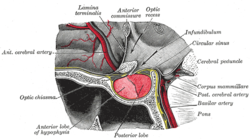
Located at the base of the brain, the pituitary gland is protected by a bony structure called the sella turcica of the sphenoid bone. |
|
Median sagittal through the hypophysis of an adult monkey. Semidiagrammatic. |
|
| Details | |
| Precursor | neural and oral ectoderm, including Rathke’s pouch |
| Artery | superior hypophyseal artery, infundibular artery, prechiasmal artery, inferior hypophyseal artery, capsular artery, artery of the inferior cavernous sinus[1] |
| Identifiers | |
| Latin | hypophysis, glandula pituitaria |
| MeSH | D010902 |
| NeuroLex ID | birnlex_1353 |
| TA98 | A11.1.00.001 |
| TA2 | 3853 |
| FMA | 13889 |
| Anatomical terms of neuroanatomy
[edit on Wikidata] |
An explanation of the development of the pituitary gland (Hypophysis cerebri) & the congenital anomalies.

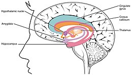
Location of the human hypothalamus.
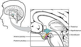
The Hypothalamus-Pituitary Complex.
In vertebrate anatomy, the pituitary gland, or hypophysis, is an endocrine gland, about the size of a chickpea[2] and weighing, on average, 0.5 grams (0.018 oz) in humans. It is a protrusion off the bottom of the hypothalamus at the base of the brain. The hypophysis rests upon the hypophyseal fossa of the sphenoid bone in the center of the middle cranial fossa and is surrounded by a small bony cavity (sella turcica) covered by a dural fold (diaphragma sellae).[3]
The anterior pituitary (or adenohypophysis) is a lobe of the gland that regulates several physiological processes including stress (by secreting ACTH), growth (by secreting GH), reproduction (by secreting FSH and LH), metabolism rate (by secreting TSH) and lactation (by secreting prolactin). The intermediate lobe synthesizes and secretes melanocyte-stimulating hormone. The posterior pituitary (or neurohypophysis) is a lobe of the gland that is functionally connected to the hypothalamus by the median eminence via a small tube called the pituitary stalk (also called the infundibular stalk or the infundibulum). It regulates hydroelectrolytic stability (by secreting ADH), uterine contraction during labor and human attachment (by secreting oxytocin).
Hormones secreted from the pituitary gland help to control growth, blood pressure, energy management, all functions of the sex organs, thyroid glands and metabolism as well as some aspects of pregnancy, childbirth, breastfeeding, water/salt concentration at the kidneys, temperature regulation and pain relief.
Structure[edit]
The pituitary gland, in humans, is oval in shape and is a pea-sized gland that sits in a protective bony enclosure called the sella turcica. It is composed of two lobes: anterior and posterior, with the intermediate lobe that joins the two regions.[4] In many animals, these three lobes are distinct. The intermediate is avascular and almost absent in human beings. The intermediate lobe is present in many animal species, in particular in rodents, mice and rats, that have been used extensively to study pituitary development and function.[5] In all animals, the fleshy, glandular anterior pituitary is distinct from the neural composition of the posterior pituitary, which is an extension of the hypothalamus.[5]

Histology of pituitary gland
The height of the pituitary gland ranges from 5.3 to 7.0 mm. The volume of the pituitary gland ranges from 200 to 440 mm3.[6]
Anterior[edit]
The anterior pituitary arises from an invagination of the oral ectoderm (Rathke’s pouch). This contrasts with the posterior pituitary, which originates from neuroectoderm.
Endocrine cells of the anterior pituitary are controlled by regulatory hormones released by parvocellular neurosecretory cells in the hypothalamic capillaries leading to infundibular blood vessels, which in turn lead to a second capillary bed in the anterior pituitary. This vascular relationship constitutes the hypothalamo-hypophyseal portal system. Diffusing out of the second capillary bed, the hypothalamic releasing hormones then bind to anterior pituitary endocrine cells, upregulating or downregulating their release of hormones.[7]
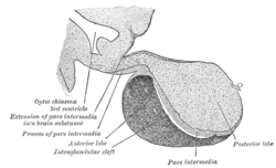
The anterior lobe of the pituitary can be divided into the pars tuberalis (pars infundibularis) and pars distalis (pars glandularis) that constitutes ~80% of the gland. The pars intermedia (the intermediate lobe) lies between the pars distalis and the pars tuberalis, and is rudimentary in the human, although in other species it is more developed.[5] It develops from a depression in the dorsal wall of the pharynx (stomal part) known as Rathke’s pouch.
The anterior pituitary contains several different types of cells[8] that synthesize and secrete hormones. Usually there is one type of cell for each major hormone formed in anterior pituitary. With special stains attached to high-affinity antibodies that bind with distinctive hormone, at least 5 types of cells can be differentiated.
| S.No. | Type of cell | Hormone secreted | Percentage of type of cell |
|---|---|---|---|
| 1. | Somatotropes | human Growth Hormone (hGH) | 30-50% |
| 2. | Corticotropes | AdrenoCorticoTropic Hormone (ACTH) | 20% |
| 3. | Thyrotropes | Thyroid-Stimulating Hormone (TSH) | 3–5% |
| 4. | Gonadotropes | Gonadotropic hormones = both Luteinizing Hormone (LH) and Follicle-Stimulating Hormone (FSH) | 3–5% |
| 5. | Lactotropes | Prolactin (PRL) | 3–5% |
Posterior[edit]
The posterior lobe develops as an extension of the hypothalamus, from the floor of the third ventricle. The posterior pituitary hormones are synthesized by cell bodies in the hypothalamus. The magnocellular neurosecretory cells, of the supraoptic and paraventricular nuclei located in the hypothalamus, project axons down the infundibulum to terminals in the posterior pituitary. This simple arrangement differs sharply from that of the adjacent anterior pituitary, which does not develop from the hypothalamus.
The release of pituitary hormones by both the anterior and posterior lobes is under the control of the hypothalamus, albeit in different ways.[7]
Functions[edit]

Anterior[edit]
The anterior pituitary synthesizes and secretes hormones. All releasing hormones (-RH) referred to, can also be referred to as releasing factors (-RF).
Somatotropes:
- Human growth hormone (HGH), also referred to as ‘growth hormone’ (GH), and also as somatotropin, is released under the influence of hypothalamic growth hormone-releasing hormone (GHRH), and is inhibited by hypothalamic somatostatin.
Corticotropes:
- Cleaved from the precursor proopiomelanocortin protein, and include adrenocorticotropic hormone (ACTH), and beta-endorphin, and melanocyte-stimulating hormone are released under the influence of hypothalamic corticotropin-releasing hormone (CRH).[9][10]: 1210
Thyrotropes:
- Thyroid-stimulating hormone (TSH), is released under the influence of hypothalamic thyrotropin-releasing hormone (TRH) and is inhibited by somatostatin.
Gonadotropes:
- Luteinizing hormone (LH).
- Follicle-stimulating hormone (FSH), both released under influence of Gonadotropin-releasing Hormone (GnRH)
Lactotropes:
- Prolactin (PRL), whose release is inconsistently stimulated by hypothalamic TRH, oxytocin, vasopressin, vasoactive intestinal peptide, angiotensin II, neuropeptide Y, galanin, substance P, bombesin-like peptides (gastrin-releasing peptide, neuromedin B and C), and neurotensin, and inhibited by hypothalamic dopamine.[11]
These hormones are released from the anterior pituitary under the influence of the hypothalamus. Hypothalamic hormones are secreted to the anterior lobe by way of a special capillary system, called the hypothalamic-hypophysial portal system.
There is also a non-endocrine cell population called folliculostellate cells.
Intermediate[edit]
The intermediate lobe synthesizes and secretes the following important endocrine hormone:
- Melanocyte–stimulating hormone (MSH). This is also produced in the anterior lobe.[12] When produced in the intermediate lobe, MSHs are sometimes called «intermedins».
Posterior[edit]
The posterior pituitary stores and secretes (but does not synthesize) the following important endocrine hormones:
Magnocellular neurons:
- Antidiuretic hormone (ADH, also known as vasopressin and arginine vasopressin AVP), the majority of which is released from the supraoptic nucleus in the hypothalamus.
- Oxytocin, most of which is released from the paraventricular nucleus in the hypothalamus. Oxytocin is one of the few hormones to create a positive feedback loop. For example, uterine contractions stimulate the release of oxytocin from the posterior pituitary, which, in turn, increases uterine contractions. This positive feedback loop continues throughout labour.
Hormones[edit]
Hormones secreted from the pituitary gland help control the following body processes:
- Growth (GH)
- Blood pressure
- Some aspects of pregnancy and childbirth including stimulation of uterine contractions
- Breast milk production
- Sex organ functions in both sexes
- Thyroid gland function
- Metabolic conversion of food into energy
- Water and osmolarity regulation in the body
- Water balance via the control of reabsorption of water by the kidneys
- Temperature regulation
- Pain relief
Clinical significance[edit]

A normal-sized hand (left) and the enlarged hand of someone with acromegaly (right)
Some of the diseases involving the pituitary gland are:
- Central diabetes insipidus caused by a deficiency of vasopressin
- Gigantism and acromegaly caused by an excess of growth hormone in childhood and adult, respectively
- Hypothyroidism caused by a deficiency of thyroid-stimulating hormone
- Hyperpituitarism, the increased (hyper) secretion of one or more of the hormones normally produced by the pituitary gland
- Hypopituitarism, the decreased (hypo) secretion of one or more of the hormones normally produced by the pituitary gland
- Panhypopituitarism a decreased secretion of most of the pituitary hormones
- Pituitary tumours
- Pituitary adenomas, noncancerous tumors that occur in the pituitary gland
All of the functions of the pituitary gland can be adversely affected by an over- or under-production of associated hormones.
The pituitary gland is important for mediating the stress response, via the hypothalamic–pituitary–adrenal axis (HPA axis) Critically, pituitary gland growth during adolescence can be altered by early life stress such as childhood maltreatment or maternal dysphoric behavior.[13]
It has been demonstrated that, after controlling for age, sex, and BMI, larger quantities of DHEA and DHEA-S tended to be linked to larger pituitary volume.[14] Additionally, a correlation between pituitary gland volume and Social Anxiety subscale scores was identified which provided a basis for exploring mediation. Again controlling for age, sex, and BMI, DHEA and DHEA-S have been found to be predictive of larger pituitary gland volume, which was also associated with increased ratings of social anxiety.[14] This research provides evidence that pituitary gland volume mediates the link between higher DHEA(S) levels (associated with relatively early adrenarche) and traits associated with social anxiety.[14] Children who experience early adrenarcheal development tend to have larger pituitary gland volume compared to children with later adrenarcheal development.[14]
History[edit]
Etymology[edit]
Pituitary gland[edit]
The Greek physician Galen referred to the pituitary gland by only using the (Ancient Greek) name ἀδήν,[15] gland.[16] He described the pituitary gland as part of a series of secretory organs for the excretion of nasal mucus.[15] Anatomist Andreas Vesalius translated ἀδήν with glans, in quam pituita destillat, «gland in which slime (pituita[17]) drips».[15][18] Besides this ‘descriptive’ name, Vesalius used glandula pituitaria, from which the English name pituitary gland[19] is ultimately derived.
The expression glandula pituitaria is still used as official synonym beside hypophysis in the official Latin nomenclature Terminologia Anatomica.[20] In the seventeenth century the supposed function of the pituitary gland to produce nasal mucus was debunked.[15] The expression glandula pituitaria and its English equivalent pituitary gland can only be justified from a historical point of view.[21] The inclusion of this synonym is merely justified by noting that the main term hypophysis is a much less popular term.[22]
Hypophysis[edit]
Note: hypophysial (or hypophyseal) means «related to the hypophysis (pituitary gland)».
The anatomist Samuel Thomas von Sömmerring coined the name hypophysis.[15] This name consists[15][21] of ὑπό (‘under’)[16] and φύειν (‘to grow’).[16] In later Greek ὑπόφυσις is used differently by Greek physicians as outgrowth.[15] Sömmering also used the equivalent expression appendix cerebri,[15][18] with appendix as appendage.[17] In various languages, Hirnanhang[18] in German and hersenaanhangsel[23] in Dutch, the terms are derived from appendix cerebri.
Other animals[edit]
The pituitary gland is found in all vertebrates, but its structure varies among different groups.
The division of the pituitary described above is typical of mammals, and is also true, to varying degrees, of all tetrapods. However, only in mammals does the posterior pituitary have a compact shape. In lungfish, it is a relatively flat sheet of tissue lying above the anterior pituitary, but in amphibians, reptiles, and birds, it becomes increasingly well developed. The intermediate lobe is, in general, not well developed in any species and is entirely absent in birds.[24]
The structure of the pituitary in fish, apart from the lungfish, is generally different from that in other animals. In general, the intermediate lobe tends to be well developed, and may equal the remainder of the anterior pituitary in size. The posterior lobe typically forms a sheet of tissue at the base of the pituitary stalk, and in most cases sends irregular finger-like projection into the tissue of the anterior pituitary, which lies directly beneath it. The anterior pituitary is typically divided into two regions, a more anterior rostral portion and a posterior proximal portion, but the boundary between the two is often not clearly marked. In elasmobranchs there is an additional, ventral lobe beneath the anterior pituitary proper.[24]
The arrangement in lampreys, which are among the most primitive of all fish, may indicate how the pituitary originally evolved in ancestral vertebrates. Here, the posterior pituitary is a simple flat sheet of tissue at the base of the brain, and there is no pituitary stalk. Rathke’s pouch remains open to the outside, close to the nasal openings. Closely associated with the pouch are three distinct clusters of glandular tissue, corresponding to the intermediate lobe, and the rostral and proximal portions of the anterior pituitary. These various parts are separated by meningial membranes, suggesting that the pituitary of other vertebrates may have formed from the fusion of a pair of separate, but associated, glands.[24]
Most armadillos also possess a neural secretory gland very similar in form to the posterior pituitary, but located in the tail and associated with the spinal cord. This may have a function in osmoregulation.[24]
There is a structure analogous to the pituitary in the octopus brain.[25]
Intermediate lobe[edit]
Although rudimentary in humans (and often considered part of the anterior pituitary), the intermediate lobe located between the anterior and posterior pituitary is important to many animals. For instance, in fish, it is believed to control physiological color change. In adult humans, it is just a thin layer of cells between the anterior and posterior pituitary. The intermediate lobe produces melanocyte-stimulating hormone (MSH), although this function is often (imprecisely) attributed to the anterior pituitary.
The intermediate lobe is, in general, not well developed in tetrapods, and is entirely absent in birds.[24]
-

Location of the pituitary gland in the human brain
-

Pituitary and pineal glands
-

The arteries of the base of the brain.
-

Mesal aspect of a brain sectioned in the median sagittal plane.
-

Pituitary
-

Pituitary gland
-
Cerebrum.Inferior view.Deep dissection.


See also[edit]
- Head and neck anatomy
- Chromophobe cell
-
- Melanotroph
- Chromophil
-
- Acidophil cell
- Basophil cell
- Oxyphil cell (parathyroid)
- Neuroendocrine cell
References[edit]
- ^ Gibo H, Hokama M, Kyoshima K, Kobayashi S (1993). «[Arteries to the pituitary]». Nippon Rinsho. 51 (10): 2550–4. PMID 8254920.
- ^ Leng, Gareth (2018). The Heart of the Brain: The Hypothalamus and its Hormones.
The gland in humans is described in Wikipedia as being the size of a pea. So common is this description that it seemed likely to be wrong, as I confirmed by examining a selection of peas. Wikipedia also gives the weight of the human pituitary as about half a gram, and in this it is more correct. The pituitary in a human is at least five times the average size of my peas; it is more like the size of a chickpea.
- ^ Mancall, Elliott L.; Brock, David G., eds. (2011). «Cranial Fossae». Gray’s Clinical Anatomy. Elsevier Health Sciences. p. 154. ISBN 9781437735802.
- ^ Ganapathy MK, Tadi P (Jan 2020). «Anatomy, Head and Neck, Pituitary Gland». StatPearls Publishing. PMID 31855373. Retrieved 24 Sep 2020.
- ^ a b c Melmed, Shlomo (2011). The Pituitary — (Third ed.). San Diego, CA 92101-4495, USA: Academic Press is an imprint of Elsevier. pp. 23–25. ISBN 978-0-12-380926-1.
{{cite book}}: CS1 maint: location (link) - ^ Yadav, Pratiksha; Singhal, Shubham; Chauhan, Surbhi; Harit, Saumya (2017). «MRI Evaluation of Size and Shape of Normal Pituitary Gland: Age and Sex Related Changes». Journal of Clinical and Diagnostic Research. doi:10.7860/JCDR/2017/31034.10933.
- ^ a b Boron, Walter F.; Boulpaep, Emile L. (2009). Medical Physiology (2nd ed.). Philadelphia: Saunders Elsevier. pp. 1016–1017. ISBN 978-1-4160-3115-4.
- ^ Textbook of Medical Physiology. Elsevier Saunders.
- ^ Knepel, W; Homolka, L; Vlaskovska, M; Nutto, D (1984). «Stimulation of adrenocorticotropin/beta-endorphin release by synthetic ovine corticotropin-releasing factor in vitro: Enhancement by various vasopressin analogs». Neuroendocrinology. 38 (5): 344–50. doi:10.1159/000123915. PMID 6328345.
- ^ Brunton, Laurence L.; Chabner, Bruce A.; Knollmann, Björn C., eds. (2011). Goodman & Gilman’s pharmacological basis of therapeutics (12th ed.). New York: McGraw-Hill. ISBN 978-0-07-162442-8.
- ^ Shlomo Melmed (3 December 2010). The pituitary. Academic Press. p. 40. ISBN 978-0-12-380926-1.
- ^ Pocock, Gillian (2006). Human Physiology (Third ed.). Oxford University Press. p. 193. ISBN 978-0-19-856878-0.
- ^ Ganella, Despina E.; Allen, Nicholas B.; Simmons, Julian G.; Schwartz, Orli; Kim, Jee Hyun; Sheeber, Lisa; Whittle, Sarah (2015). «Early life stress alters pituitary growth during adolescence—A longitudinal study». Psychoneuroendocrinology. 53: 185–194. doi:10.1016/j.psyneuen.2015.01.005. hdl:10536/DRO/DU:30144589. PMID 25622011. S2CID 5247274.
- ^ a b c d Murray, CR; Simmons, JG; Allen, NB; Byrne, ML; Mundy, LK; Seal, ML; Patton, GC; Olsson, CA; Whittle, S (February 2016). «Associations between dehydroepiandrosterone (DHEA) levels, pituitary volume, and social anxiety in children». Psychoneuroendocrinology. 64: 31–9. doi:10.1016/j.psyneuen.2015.11.004. PMID 26600008. S2CID 22520320.
- ^ a b c d e f g h Hyrtl, J. (1880). Onomatologia Anatomica. Geschichte und Kritik der anatomischen Sprache der Gegenwart. Wien: Wilhelm Braumüller. K.K. Hof- und Universitätsbuchhändler.
- ^ a b c Liddell, H.G. & Scott, R. (1940). A Greek-English Lexicon. revised and augmented throughout by Sir Henry Stuart Jones. with the assistance of. Roderick McKenzie. Oxford: Clarendon Press.
- ^ a b Lewis, C.T. & Short, C. (1879). A Latin dictionary founded on Andrews’ edition of Freund’s Latin dictionary. Oxford: Clarendon Press.
- ^ a b c Schreger, C.H.Th.(1805). Synonymia anatomica. Synonymik der anatomischen Nomenclatur. Fürth: im Bureau für Literatur.
- ^ Anderson, D.M. (2000). Dorland’s illustrated medical dictionary (29th edition). Philadelphia/London/Toronto/Montreal/Sydney/Tokyo: W.B. Saunders Company.
- ^ Federative Committee on Anatomical Terminology (FCAT) (1998). Terminologia Anatomica. Stuttgart: Thieme
- ^ a b Triepel, H. (1927). Die anatomischen Namen. Ihre Ableitung und Aussprache. Anhang: Biographische Notizen.(Elfte Auflage). München: Verlag J.F. Bergmann.
- ^ International Anatomical Nomenclature Committee (1966). Nomina Anatomica. Amsterdam: Excerpta Medica Foundation, p. 62
- ^ Pinkhof, H. (1923). Vertalend en verklarend woordenboek van uitheemsche geneeskundige termen. Haarlem: De Erven F. Bohn.
- ^ a b c d e Romer, Alfred Sherwood; Parsons, Thomas S. (1977). The Vertebrate Body. Philadelphia, PA: Holt-Saunders International. pp. 549–550. ISBN 0-03-910284-X.
- ^ Wells, M. J.; Wells, J. (1969). «Pituitary Analogue in the Octopus». Nature. 222 (5190): 293–294. Bibcode:1969Natur.222..293W. doi:10.1038/222293a0. PMID 5778406. S2CID 4159935.
External links[edit]
- hier-382 at NeuroNames
- Histology image: 14201loa – Histology Learning System at Boston University
- The Pituitary Gland, from the UMM Endocrinology Health Guide Archived 2010-06-20 at the Wayback Machine
- Oklahoma State, Endocrine System
- The Pituitary Foundation
- The Pituitary Network Association — pituitary.org
From Wikipedia, the free encyclopedia
| Pituitary gland | |
|---|---|
|
Located at the base of the brain, the pituitary gland is protected by a bony structure called the sella turcica of the sphenoid bone. |
|
|
Median sagittal through the hypophysis of an adult monkey. Semidiagrammatic. |
|
| Details | |
| Precursor | neural and oral ectoderm, including Rathke’s pouch |
| Artery | superior hypophyseal artery, infundibular artery, prechiasmal artery, inferior hypophyseal artery, capsular artery, artery of the inferior cavernous sinus[1] |
| Identifiers | |
| Latin | hypophysis, glandula pituitaria |
| MeSH | D010902 |
| NeuroLex ID | birnlex_1353 |
| TA98 | A11.1.00.001 |
| TA2 | 3853 |
| FMA | 13889 |
| Anatomical terms of neuroanatomy
[edit on Wikidata] |
An explanation of the development of the pituitary gland (Hypophysis cerebri) & the congenital anomalies.
Location of the human hypothalamus.
The Hypothalamus-Pituitary Complex.
In vertebrate anatomy, the pituitary gland, or hypophysis, is an endocrine gland, about the size of a chickpea[2] and weighing, on average, 0.5 grams (0.018 oz) in humans. It is a protrusion off the bottom of the hypothalamus at the base of the brain. The hypophysis rests upon the hypophyseal fossa of the sphenoid bone in the center of the middle cranial fossa and is surrounded by a small bony cavity (sella turcica) covered by a dural fold (diaphragma sellae).[3]
The anterior pituitary (or adenohypophysis) is a lobe of the gland that regulates several physiological processes including stress (by secreting ACTH), growth (by secreting GH), reproduction (by secreting FSH and LH), metabolism rate (by secreting TSH) and lactation (by secreting prolactin). The intermediate lobe synthesizes and secretes melanocyte-stimulating hormone. The posterior pituitary (or neurohypophysis) is a lobe of the gland that is functionally connected to the hypothalamus by the median eminence via a small tube called the pituitary stalk (also called the infundibular stalk or the infundibulum). It regulates hydroelectrolytic stability (by secreting ADH), uterine contraction during labor and human attachment (by secreting oxytocin).
Hormones secreted from the pituitary gland help to control growth, blood pressure, energy management, all functions of the sex organs, thyroid glands and metabolism as well as some aspects of pregnancy, childbirth, breastfeeding, water/salt concentration at the kidneys, temperature regulation and pain relief.
Structure[edit]
The pituitary gland, in humans, is oval in shape and is a pea-sized gland that sits in a protective bony enclosure called the sella turcica. It is composed of two lobes: anterior and posterior, with the intermediate lobe that joins the two regions.[4] In many animals, these three lobes are distinct. The intermediate is avascular and almost absent in human beings. The intermediate lobe is present in many animal species, in particular in rodents, mice and rats, that have been used extensively to study pituitary development and function.[5] In all animals, the fleshy, glandular anterior pituitary is distinct from the neural composition of the posterior pituitary, which is an extension of the hypothalamus.[5]
Histology of pituitary gland
The height of the pituitary gland ranges from 5.3 to 7.0 mm. The volume of the pituitary gland ranges from 200 to 440 mm3.[6]
Anterior[edit]
The anterior pituitary arises from an invagination of the oral ectoderm (Rathke’s pouch). This contrasts with the posterior pituitary, which originates from neuroectoderm.
Endocrine cells of the anterior pituitary are controlled by regulatory hormones released by parvocellular neurosecretory cells in the hypothalamic capillaries leading to infundibular blood vessels, which in turn lead to a second capillary bed in the anterior pituitary. This vascular relationship constitutes the hypothalamo-hypophyseal portal system. Diffusing out of the second capillary bed, the hypothalamic releasing hormones then bind to anterior pituitary endocrine cells, upregulating or downregulating their release of hormones.[7]
The anterior lobe of the pituitary can be divided into the pars tuberalis (pars infundibularis) and pars distalis (pars glandularis) that constitutes ~80% of the gland. The pars intermedia (the intermediate lobe) lies between the pars distalis and the pars tuberalis, and is rudimentary in the human, although in other species it is more developed.[5] It develops from a depression in the dorsal wall of the pharynx (stomal part) known as Rathke’s pouch.
The anterior pituitary contains several different types of cells[8] that synthesize and secrete hormones. Usually there is one type of cell for each major hormone formed in anterior pituitary. With special stains attached to high-affinity antibodies that bind with distinctive hormone, at least 5 types of cells can be differentiated.
| S.No. | Type of cell | Hormone secreted | Percentage of type of cell |
|---|---|---|---|
| 1. | Somatotropes | human Growth Hormone (hGH) | 30-50% |
| 2. | Corticotropes | AdrenoCorticoTropic Hormone (ACTH) | 20% |
| 3. | Thyrotropes | Thyroid-Stimulating Hormone (TSH) | 3–5% |
| 4. | Gonadotropes | Gonadotropic hormones = both Luteinizing Hormone (LH) and Follicle-Stimulating Hormone (FSH) | 3–5% |
| 5. | Lactotropes | Prolactin (PRL) | 3–5% |
Posterior[edit]
The posterior lobe develops as an extension of the hypothalamus, from the floor of the third ventricle. The posterior pituitary hormones are synthesized by cell bodies in the hypothalamus. The magnocellular neurosecretory cells, of the supraoptic and paraventricular nuclei located in the hypothalamus, project axons down the infundibulum to terminals in the posterior pituitary. This simple arrangement differs sharply from that of the adjacent anterior pituitary, which does not develop from the hypothalamus.
The release of pituitary hormones by both the anterior and posterior lobes is under the control of the hypothalamus, albeit in different ways.[7]
Functions[edit]
Anterior[edit]
The anterior pituitary synthesizes and secretes hormones. All releasing hormones (-RH) referred to, can also be referred to as releasing factors (-RF).
Somatotropes:
- Human growth hormone (HGH), also referred to as ‘growth hormone’ (GH), and also as somatotropin, is released under the influence of hypothalamic growth hormone-releasing hormone (GHRH), and is inhibited by hypothalamic somatostatin.
Corticotropes:
- Cleaved from the precursor proopiomelanocortin protein, and include adrenocorticotropic hormone (ACTH), and beta-endorphin, and melanocyte-stimulating hormone are released under the influence of hypothalamic corticotropin-releasing hormone (CRH).[9][10]: 1210
Thyrotropes:
- Thyroid-stimulating hormone (TSH), is released under the influence of hypothalamic thyrotropin-releasing hormone (TRH) and is inhibited by somatostatin.
Gonadotropes:
- Luteinizing hormone (LH).
- Follicle-stimulating hormone (FSH), both released under influence of Gonadotropin-releasing Hormone (GnRH)
Lactotropes:
- Prolactin (PRL), whose release is inconsistently stimulated by hypothalamic TRH, oxytocin, vasopressin, vasoactive intestinal peptide, angiotensin II, neuropeptide Y, galanin, substance P, bombesin-like peptides (gastrin-releasing peptide, neuromedin B and C), and neurotensin, and inhibited by hypothalamic dopamine.[11]
These hormones are released from the anterior pituitary under the influence of the hypothalamus. Hypothalamic hormones are secreted to the anterior lobe by way of a special capillary system, called the hypothalamic-hypophysial portal system.
There is also a non-endocrine cell population called folliculostellate cells.
Intermediate[edit]
The intermediate lobe synthesizes and secretes the following important endocrine hormone:
- Melanocyte–stimulating hormone (MSH). This is also produced in the anterior lobe.[12] When produced in the intermediate lobe, MSHs are sometimes called «intermedins».
Posterior[edit]
The posterior pituitary stores and secretes (but does not synthesize) the following important endocrine hormones:
Magnocellular neurons:
- Antidiuretic hormone (ADH, also known as vasopressin and arginine vasopressin AVP), the majority of which is released from the supraoptic nucleus in the hypothalamus.
- Oxytocin, most of which is released from the paraventricular nucleus in the hypothalamus. Oxytocin is one of the few hormones to create a positive feedback loop. For example, uterine contractions stimulate the release of oxytocin from the posterior pituitary, which, in turn, increases uterine contractions. This positive feedback loop continues throughout labour.
Hormones[edit]
Hormones secreted from the pituitary gland help control the following body processes:
- Growth (GH)
- Blood pressure
- Some aspects of pregnancy and childbirth including stimulation of uterine contractions
- Breast milk production
- Sex organ functions in both sexes
- Thyroid gland function
- Metabolic conversion of food into energy
- Water and osmolarity regulation in the body
- Water balance via the control of reabsorption of water by the kidneys
- Temperature regulation
- Pain relief
Clinical significance[edit]
A normal-sized hand (left) and the enlarged hand of someone with acromegaly (right)
Some of the diseases involving the pituitary gland are:
- Central diabetes insipidus caused by a deficiency of vasopressin
- Gigantism and acromegaly caused by an excess of growth hormone in childhood and adult, respectively
- Hypothyroidism caused by a deficiency of thyroid-stimulating hormone
- Hyperpituitarism, the increased (hyper) secretion of one or more of the hormones normally produced by the pituitary gland
- Hypopituitarism, the decreased (hypo) secretion of one or more of the hormones normally produced by the pituitary gland
- Panhypopituitarism a decreased secretion of most of the pituitary hormones
- Pituitary tumours
- Pituitary adenomas, noncancerous tumors that occur in the pituitary gland
All of the functions of the pituitary gland can be adversely affected by an over- or under-production of associated hormones.
The pituitary gland is important for mediating the stress response, via the hypothalamic–pituitary–adrenal axis (HPA axis) Critically, pituitary gland growth during adolescence can be altered by early life stress such as childhood maltreatment or maternal dysphoric behavior.[13]
It has been demonstrated that, after controlling for age, sex, and BMI, larger quantities of DHEA and DHEA-S tended to be linked to larger pituitary volume.[14] Additionally, a correlation between pituitary gland volume and Social Anxiety subscale scores was identified which provided a basis for exploring mediation. Again controlling for age, sex, and BMI, DHEA and DHEA-S have been found to be predictive of larger pituitary gland volume, which was also associated with increased ratings of social anxiety.[14] This research provides evidence that pituitary gland volume mediates the link between higher DHEA(S) levels (associated with relatively early adrenarche) and traits associated with social anxiety.[14] Children who experience early adrenarcheal development tend to have larger pituitary gland volume compared to children with later adrenarcheal development.[14]
History[edit]
Etymology[edit]
Pituitary gland[edit]
The Greek physician Galen referred to the pituitary gland by only using the (Ancient Greek) name ἀδήν,[15] gland.[16] He described the pituitary gland as part of a series of secretory organs for the excretion of nasal mucus.[15] Anatomist Andreas Vesalius translated ἀδήν with glans, in quam pituita destillat, «gland in which slime (pituita[17]) drips».[15][18] Besides this ‘descriptive’ name, Vesalius used glandula pituitaria, from which the English name pituitary gland[19] is ultimately derived.
The expression glandula pituitaria is still used as official synonym beside hypophysis in the official Latin nomenclature Terminologia Anatomica.[20] In the seventeenth century the supposed function of the pituitary gland to produce nasal mucus was debunked.[15] The expression glandula pituitaria and its English equivalent pituitary gland can only be justified from a historical point of view.[21] The inclusion of this synonym is merely justified by noting that the main term hypophysis is a much less popular term.[22]
Hypophysis[edit]
Note: hypophysial (or hypophyseal) means «related to the hypophysis (pituitary gland)».
The anatomist Samuel Thomas von Sömmerring coined the name hypophysis.[15] This name consists[15][21] of ὑπό (‘under’)[16] and φύειν (‘to grow’).[16] In later Greek ὑπόφυσις is used differently by Greek physicians as outgrowth.[15] Sömmering also used the equivalent expression appendix cerebri,[15][18] with appendix as appendage.[17] In various languages, Hirnanhang[18] in German and hersenaanhangsel[23] in Dutch, the terms are derived from appendix cerebri.
Other animals[edit]
The pituitary gland is found in all vertebrates, but its structure varies among different groups.
The division of the pituitary described above is typical of mammals, and is also true, to varying degrees, of all tetrapods. However, only in mammals does the posterior pituitary have a compact shape. In lungfish, it is a relatively flat sheet of tissue lying above the anterior pituitary, but in amphibians, reptiles, and birds, it becomes increasingly well developed. The intermediate lobe is, in general, not well developed in any species and is entirely absent in birds.[24]
The structure of the pituitary in fish, apart from the lungfish, is generally different from that in other animals. In general, the intermediate lobe tends to be well developed, and may equal the remainder of the anterior pituitary in size. The posterior lobe typically forms a sheet of tissue at the base of the pituitary stalk, and in most cases sends irregular finger-like projection into the tissue of the anterior pituitary, which lies directly beneath it. The anterior pituitary is typically divided into two regions, a more anterior rostral portion and a posterior proximal portion, but the boundary between the two is often not clearly marked. In elasmobranchs there is an additional, ventral lobe beneath the anterior pituitary proper.[24]
The arrangement in lampreys, which are among the most primitive of all fish, may indicate how the pituitary originally evolved in ancestral vertebrates. Here, the posterior pituitary is a simple flat sheet of tissue at the base of the brain, and there is no pituitary stalk. Rathke’s pouch remains open to the outside, close to the nasal openings. Closely associated with the pouch are three distinct clusters of glandular tissue, corresponding to the intermediate lobe, and the rostral and proximal portions of the anterior pituitary. These various parts are separated by meningial membranes, suggesting that the pituitary of other vertebrates may have formed from the fusion of a pair of separate, but associated, glands.[24]
Most armadillos also possess a neural secretory gland very similar in form to the posterior pituitary, but located in the tail and associated with the spinal cord. This may have a function in osmoregulation.[24]
There is a structure analogous to the pituitary in the octopus brain.[25]
Intermediate lobe[edit]
Although rudimentary in humans (and often considered part of the anterior pituitary), the intermediate lobe located between the anterior and posterior pituitary is important to many animals. For instance, in fish, it is believed to control physiological color change. In adult humans, it is just a thin layer of cells between the anterior and posterior pituitary. The intermediate lobe produces melanocyte-stimulating hormone (MSH), although this function is often (imprecisely) attributed to the anterior pituitary.
The intermediate lobe is, in general, not well developed in tetrapods, and is entirely absent in birds.[24]
-
Location of the pituitary gland in the human brain
-
Pituitary and pineal glands
-
The arteries of the base of the brain.
-
Mesal aspect of a brain sectioned in the median sagittal plane.
-
Pituitary
-
Pituitary gland
-
Cerebrum.Inferior view.Deep dissection.
See also[edit]
- Head and neck anatomy
- Chromophobe cell
-
- Melanotroph
- Chromophil
-
- Acidophil cell
- Basophil cell
- Oxyphil cell (parathyroid)
- Neuroendocrine cell
References[edit]
- ^ Gibo H, Hokama M, Kyoshima K, Kobayashi S (1993). «[Arteries to the pituitary]». Nippon Rinsho. 51 (10): 2550–4. PMID 8254920.
- ^ Leng, Gareth (2018). The Heart of the Brain: The Hypothalamus and its Hormones.
The gland in humans is described in Wikipedia as being the size of a pea. So common is this description that it seemed likely to be wrong, as I confirmed by examining a selection of peas. Wikipedia also gives the weight of the human pituitary as about half a gram, and in this it is more correct. The pituitary in a human is at least five times the average size of my peas; it is more like the size of a chickpea.
- ^ Mancall, Elliott L.; Brock, David G., eds. (2011). «Cranial Fossae». Gray’s Clinical Anatomy. Elsevier Health Sciences. p. 154. ISBN 9781437735802.
- ^ Ganapathy MK, Tadi P (Jan 2020). «Anatomy, Head and Neck, Pituitary Gland». StatPearls Publishing. PMID 31855373. Retrieved 24 Sep 2020.
- ^ a b c Melmed, Shlomo (2011). The Pituitary — (Third ed.). San Diego, CA 92101-4495, USA: Academic Press is an imprint of Elsevier. pp. 23–25. ISBN 978-0-12-380926-1.
{{cite book}}: CS1 maint: location (link) - ^ Yadav, Pratiksha; Singhal, Shubham; Chauhan, Surbhi; Harit, Saumya (2017). «MRI Evaluation of Size and Shape of Normal Pituitary Gland: Age and Sex Related Changes». Journal of Clinical and Diagnostic Research. doi:10.7860/JCDR/2017/31034.10933.
- ^ a b Boron, Walter F.; Boulpaep, Emile L. (2009). Medical Physiology (2nd ed.). Philadelphia: Saunders Elsevier. pp. 1016–1017. ISBN 978-1-4160-3115-4.
- ^ Textbook of Medical Physiology. Elsevier Saunders.
- ^ Knepel, W; Homolka, L; Vlaskovska, M; Nutto, D (1984). «Stimulation of adrenocorticotropin/beta-endorphin release by synthetic ovine corticotropin-releasing factor in vitro: Enhancement by various vasopressin analogs». Neuroendocrinology. 38 (5): 344–50. doi:10.1159/000123915. PMID 6328345.
- ^ Brunton, Laurence L.; Chabner, Bruce A.; Knollmann, Björn C., eds. (2011). Goodman & Gilman’s pharmacological basis of therapeutics (12th ed.). New York: McGraw-Hill. ISBN 978-0-07-162442-8.
- ^ Shlomo Melmed (3 December 2010). The pituitary. Academic Press. p. 40. ISBN 978-0-12-380926-1.
- ^ Pocock, Gillian (2006). Human Physiology (Third ed.). Oxford University Press. p. 193. ISBN 978-0-19-856878-0.
- ^ Ganella, Despina E.; Allen, Nicholas B.; Simmons, Julian G.; Schwartz, Orli; Kim, Jee Hyun; Sheeber, Lisa; Whittle, Sarah (2015). «Early life stress alters pituitary growth during adolescence—A longitudinal study». Psychoneuroendocrinology. 53: 185–194. doi:10.1016/j.psyneuen.2015.01.005. hdl:10536/DRO/DU:30144589. PMID 25622011. S2CID 5247274.
- ^ a b c d Murray, CR; Simmons, JG; Allen, NB; Byrne, ML; Mundy, LK; Seal, ML; Patton, GC; Olsson, CA; Whittle, S (February 2016). «Associations between dehydroepiandrosterone (DHEA) levels, pituitary volume, and social anxiety in children». Psychoneuroendocrinology. 64: 31–9. doi:10.1016/j.psyneuen.2015.11.004. PMID 26600008. S2CID 22520320.
- ^ a b c d e f g h Hyrtl, J. (1880). Onomatologia Anatomica. Geschichte und Kritik der anatomischen Sprache der Gegenwart. Wien: Wilhelm Braumüller. K.K. Hof- und Universitätsbuchhändler.
- ^ a b c Liddell, H.G. & Scott, R. (1940). A Greek-English Lexicon. revised and augmented throughout by Sir Henry Stuart Jones. with the assistance of. Roderick McKenzie. Oxford: Clarendon Press.
- ^ a b Lewis, C.T. & Short, C. (1879). A Latin dictionary founded on Andrews’ edition of Freund’s Latin dictionary. Oxford: Clarendon Press.
- ^ a b c Schreger, C.H.Th.(1805). Synonymia anatomica. Synonymik der anatomischen Nomenclatur. Fürth: im Bureau für Literatur.
- ^ Anderson, D.M. (2000). Dorland’s illustrated medical dictionary (29th edition). Philadelphia/London/Toronto/Montreal/Sydney/Tokyo: W.B. Saunders Company.
- ^ Federative Committee on Anatomical Terminology (FCAT) (1998). Terminologia Anatomica. Stuttgart: Thieme
- ^ a b Triepel, H. (1927). Die anatomischen Namen. Ihre Ableitung und Aussprache. Anhang: Biographische Notizen.(Elfte Auflage). München: Verlag J.F. Bergmann.
- ^ International Anatomical Nomenclature Committee (1966). Nomina Anatomica. Amsterdam: Excerpta Medica Foundation, p. 62
- ^ Pinkhof, H. (1923). Vertalend en verklarend woordenboek van uitheemsche geneeskundige termen. Haarlem: De Erven F. Bohn.
- ^ a b c d e Romer, Alfred Sherwood; Parsons, Thomas S. (1977). The Vertebrate Body. Philadelphia, PA: Holt-Saunders International. pp. 549–550. ISBN 0-03-910284-X.
- ^ Wells, M. J.; Wells, J. (1969). «Pituitary Analogue in the Octopus». Nature. 222 (5190): 293–294. Bibcode:1969Natur.222..293W. doi:10.1038/222293a0. PMID 5778406. S2CID 4159935.
External links[edit]
- hier-382 at NeuroNames
- Histology image: 14201loa – Histology Learning System at Boston University
- The Pituitary Gland, from the UMM Endocrinology Health Guide Archived 2010-06-20 at the Wayback Machine
- Oklahoma State, Endocrine System
- The Pituitary Foundation
- The Pituitary Network Association — pituitary.org
Русский[править]
Морфологические и синтаксические свойства[править]
| падеж | ед. ч. | мн. ч. |
|---|---|---|
| Им. | гипо́физ | гипо́физы |
| Р. | гипо́физа | гипо́физов |
| Д. | гипо́физу | гипо́физам |
| В. | гипо́физ | гипо́физы |
| Тв. | гипо́физом | гипо́физами |
| Пр. | гипо́физе | гипо́физах |
ги—по́—физ
Существительное, неодушевлённое, мужской род, 2-е склонение (тип склонения 1a по классификации А. А. Зализняка).
Корень: —.
Произношение[править]
- МФА: [ɡʲɪˈpofʲɪs]
Семантические свойства[править]

Значение[править]
- анат. мозговой придаток в форме округлого образования, расположенного на нижней поверхности головного мозга в костном кармане (турецком седле), гормоны которого влияют на рост, обмен веществ и репродуктивную функцию и взаимодействующий с гипоталамусом ◆ Имплантация изотопа не вызывала недостаточности надпочечников, как эктомия, и других эндокринных желез, что отмечали при тотальной эктомии гипофиза. , Медицинский реферативный журнал г. // «1969»
Синонимы[править]
- нижний мозговой придаток, питуитарная железа
Антонимы[править]
- —
Гиперонимы[править]
- эндокринная железа
Гипонимы[править]
- —
Родственные слова[править]
| Ближайшее родство | |
|
Этимология[править]
От совр. лат. hypophysis «придаток», далее из др.-греч. ὑπόφυσις «отросток», далее из ὑπό- + -φυσις;
- первая часть — из ὑπό «под, ниже»;
- вторая часть — из φύσις «вещество; природа, характер», далее из φύω «рождать, создавать», далее из праиндоевр. *bheu- «существовать, расти».
Фразеологизмы и устойчивые сочетания[править]
Перевод[править]
| Список переводов | |
|
Библиография[править]
Башкирский[править]
Морфологические и синтаксические свойства[править]
гипофиз
Существительное.
Корень: —.
Произношение[править]
Семантические свойства[править]
Значение[править]
- анат. гипофиз (аналогично русскому слову), питуитарная железа ◆ Отсутствует пример употребления (см. рекомендации).
Синонимы[править]
- ?
Антонимы[править]
- —
Гиперонимы[править]
- ?
Гипонимы[править]
- ?
Родственные слова[править]
| Ближайшее родство | |
Этимология[править]
Из ??
Фразеологизмы и устойчивые сочетания[править]
Библиография[править]
Казахский[править]
Морфологические и синтаксические свойства[править]
гипофиз
Существительное.
Корень: —.
Произношение[править]
Семантические свойства[править]
Значение[править]
- анат. гипофиз (аналогично русскому слову), питуитарная железа ◆ Отсутствует пример употребления (см. рекомендации).
Синонимы[править]
- ?
Антонимы[править]
- —
Гиперонимы[править]
- ?
Гипонимы[править]
- ?
Родственные слова[править]
| Ближайшее родство | |
Этимология[править]
Из ??
Фразеологизмы и устойчивые сочетания[править]
Библиография[править]
Киргизский[править]
Морфологические и синтаксические свойства[править]
гипофиз
Существительное.
Корень: —.
Произношение[править]
Семантические свойства[править]
Значение[править]
- анат. гипофиз (аналогично русскому слову), питуитарная железа ◆ Отсутствует пример употребления (см. рекомендации).
Синонимы[править]
- ?
Антонимы[править]
- —
Гиперонимы[править]
- ?
Гипонимы[править]
- ?
Родственные слова[править]
| Ближайшее родство | |
Этимология[править]
Из ??
Фразеологизмы и устойчивые сочетания[править]
Библиография[править]
Таджикский[править]
Морфологические и синтаксические свойства[править]
гипофиз
Существительное.
Корень: —.
Произношение[править]
Семантические свойства[править]
Значение[править]
- анат. гипофиз (аналогично русскому слову), питуитарная железа ◆ Отсутствует пример употребления (см. рекомендации).
Синонимы[править]
- ?
Антонимы[править]
- —
Гиперонимы[править]
- ?
Гипонимы[править]
- ?
Родственные слова[править]
| Ближайшее родство | |
Этимология[править]
Из ??
Фразеологизмы и устойчивые сочетания[править]
Библиография[править]
Татарский[править]
Морфологические и синтаксические свойства[править]
гипофиз
Существительное.
Корень: —.
Произношение[править]
Семантические свойства[править]
Значение[править]
- анат. гипофиз (аналогично русскому слову), питуитарная железа ◆ Отсутствует пример употребления (см. рекомендации).
Синонимы[править]
- ?
Антонимы[править]
- —
Гиперонимы[править]
- ?
Гипонимы[править]
- ?
Родственные слова[править]
| Ближайшее родство | |
Этимология[править]
Из ??
Фразеологизмы и устойчивые сочетания[править]
Библиография[править]
Чеченский[править]
Морфологические и синтаксические свойства[править]
гипофиз
Существительное.
Корень: —.
Произношение[править]
Семантические свойства[править]
Значение[править]
- анат. гипофиз (аналогично русскому слову), питуитарная железа ◆ Отсутствует пример употребления (см. рекомендации).
Синонимы[править]
- ?
Антонимы[править]
- —
Гиперонимы[править]
- ?
Гипонимы[править]
- ?
Родственные слова[править]
| Ближайшее родство | |
Этимология[править]
Из ??
Фразеологизмы и устойчивые сочетания[править]
Библиография[править]
гипофиз
→
гипофиз — существительное, именительный п., муж. p., ед. ч.
↳
гипофиз — существительное, винительный п., муж. p., ед. ч.
Часть речи: существительное
| Единственное число | Множественное число | |
|---|---|---|
| Им. |
гипофиз |
гипофизы |
| Рд. |
гипофиза |
гипофизов |
| Дт. |
гипофизу |
гипофизам |
| Вн. |
гипофиз |
гипофизы |
| Тв. |
гипофизом |
гипофизами |
| Пр. |
гипофизе |
гипофизах |
Если вы нашли ошибку, пожалуйста, выделите фрагмент текста и нажмите Ctrl+Enter.