НЕРВ ЛИЦЕВОЙ
- НЕРВ ЛИЦЕВОЙ
- (facial nerve) — седьмая пара черепных нервов (VII): смешанный чувствительный и двигательный нерв, иннервирующий мимические мышцы, вкусовые сосочки в передних двух третях языка, а также подъязычные, поднижнечелюстные и слезные железы (парасимпатическая часть -ред.). Небольшая ветвь, идущая к среднему уху, иннервирует стременную мышцу, осуществляющую движения стремени (слуховая косточка).
Толковый словарь по медицине.
2013.
Смотреть что такое «НЕРВ ЛИЦЕВОЙ» в других словарях:
-
нерв лицевой — (n. facialls) VII пара черепных нервов. Выходит из мозга у заднего края моста в мостомоз жечковом углу, рядом с промежуточным нервом, который присоединяется к лицевому нерву, являясь его составной частью. Через внутренний слуховой проход… … Словарь терминов и понятий по анатомии человека
-
Нерв Лицевой (Facial Nerve) — седьмая пара черепных нервов (VII): смешанный чувствительный и двигательный нерв, иннервирующий мимические мышцы, вкусовые сосочки в передних двух третях языка, а также подъязычные, поднижнечелюстные и слезные железы (парасимпатическая часть… … Медицинские термины
-
нерв лицевой — (n. facialis, PNA, BNA, JNA) см. Перечень анат. терминов … Большой медицинский словарь
-
Лицевой нерв — Лицевой нерв … Википедия
-
Лицевой нерв (nervus facialis) — Вид справа. лицевой нерв; височные ветви; скуловые ветви; подглазничный нерв; подбородочный нерв; щечные ветви; краевая ветвь нижней челюсти; поверхностная шейная петля; поперечный нерв шеи; шейная ветвь; большой ушной нерв; околоушная слюнная… … Атлас анатомии человека
-
ЛИЦЕВОЙ НЕРВ — (nervus facialis), VII пара черепномозговых нервов; смешанный нерв. .(Источник: «Биологический энциклопедический словарь.» Гл. ред. М. С. Гиляров; Редкол.: А. А. Бабаев, Г. Г. Винберг, Г. А. Заварзин и др. 2 е изд., исправл. М.: Сов. Энциклопедия … Биологический энциклопедический словарь
-
ЛИЦЕВОЙ НЕРВ — седьмая пара черепных нервов у позвоночных животных и человека; содержит двигательные и чувствительные нервные волокна. У человека иннервирует мимическую мускулатуру, слезные и слюнные железы, слизистую оболочку языка, неба, полости носа и… … Большой Энциклопедический словарь
-
лицевой нерв — седьмая пара черепных нервов у позвоночных животных и человека; содержит двигательные и чувствительные нервные волокна. У человека иннервирует мимическую мускулатуру, слёзные и слюнные железы, слизистую оболочку языка, нёба, полости носа и… … Энциклопедический словарь
-
лицевой — прил., употр. сравн. часто 1. Лицевым называется что либо, связанное с лицом как передней частью головы человека. Лицевой нерв. | Лицевые мышцы. 2. Лицевой стороной называется передняя, обращённая наружу часть чего либо, в противоположность… … Толковый словарь Дмитриева
-
лицевой — а/я, о/е 1) Относящийся к лицу, находящийся на лице. Лицевой нерв. Лицевые мышцы. 2) Передняя часть, передняя сторона чего л. Лицевая сторона ограды. За поселком был мост через реку Демена, после которого справа тянулась деревня Павлиново,… … Популярный словарь русского языка
From Wikipedia, the free encyclopedia
Not to be confused with other nerves that innervate the face, such as the trigeminal nerve.
| Facial nerve | |
|---|---|

The course of the facial nerve is shown here |
|
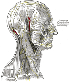
The nerves of the scalp, face, and side of neck. |
|
| Details | |
| From | facial nerve nucleus, intermediate nerve |
| To | greater superficial petrosal nerve, |
| Innervates | Motor: Muscles of facial expression, posterior belly of digastric, stylohyoid, stapedius Special sensory: taste to anterior two-thirds of tongue Parasympathetic: submandibular gland, sublingual gland, lacrimal glands |
| Identifiers | |
| Latin | nervus facialis |
| MeSH | D005154 |
| NeuroNames | 551 |
| TA98 | A14.2.01.099 |
| TA2 | 6284 |
| FMA | 50868 |
| Anatomical terms of neuroanatomy
[edit on Wikidata] |
The facial nerve, also known as the seventh cranial nerve, cranial nerve VII, or simply CN VII, is a cranial nerve that emerges from the pons of the brainstem, controls the muscles of facial expression, and functions in the conveyance of taste sensations from the anterior two-thirds of the tongue.[1][2] The nerve typically travels from the pons through the facial canal in the temporal bone and exits the skull at the stylomastoid foramen.[3] It arises from the brainstem from an area posterior to the cranial nerve VI (abducens nerve) and anterior to cranial nerve VIII (vestibulocochlear nerve).
The facial nerve also supplies preganglionic parasympathetic fibers to several head and neck ganglia.
The facial and intermediate nerves can be collectively referred to as the nervus intermediofacialis.
Structure[edit]
The path of the facial nerve can be divided into six segments:
- intracranial (cisternal) segment
- meatal (canalicular) segment (within the internal auditory canal)
- labyrinthine segment (internal auditory canal to geniculate ganglion)
- tympanic segment (from geniculate ganglion to pyramidal eminence)
- mastoid segment (from pyramidal eminence to stylomastoid foramen)
- extratemporal segment (from stylomastoid foramen to post parotid branches)
The motor part of the facial nerve arises from the facial nerve nucleus in the pons, while the sensory and parasympathetic parts of the facial nerve arise from the intermediate nerve.
From the brain stem, the motor and sensory parts of the facial nerve join together and traverse the posterior cranial fossa before entering the petrous temporal bone via the internal auditory meatus. Upon exiting the internal auditory meatus, the nerve then runs a tortuous course through the facial canal, which is divided into the labyrinthine, tympanic, and mastoid segments.
The labyrinthine segment is very short, and ends where the facial nerve forms a bend known as the geniculum of the facial nerve (genu meaning knee), which contains the geniculate ganglion for sensory nerve bodies. The first branch of the facial nerve, the greater petrosal nerve, arises here from the geniculate ganglion. The greater petrosal nerve runs through the pterygoid canal and synapses at the pterygopalatine ganglion. Postsynaptic fibers of the greater petrosal nerve innervate the lacrimal gland.
In the tympanic segment, the facial nerve runs through the tympanic cavity, medial to the incus.
The pyramidal eminence is the second bend in the facial nerve, where the nerve runs downward as the mastoid segment. In the temporal part of the facial canal, the nerve gives rise to the nerve to the stapedius muscle and chorda tympani. The chorda tympani supplies taste fibers to the anterior two thirds of the tongue, and also synapses with the submandibular ganglion. Postsynaptic fibers from the submandibular ganglion supply the sublingual and submandibular glands.
Upon emerging from the stylomastoid foramen, the facial nerve gives rise to the posterior auricular branch. The facial nerve then passes through the parotid gland, which it does not innervate, to form the parotid plexus, which splits into five branches (temporal, zygomatic, buccal, marginal mandibular, and cervical) innervating the muscles of facial expression.[4][5]
Intracranial branches[edit]
The greater petrosal nerve arises at the superior salivatory nucleus of the pons and provides parasympathetic innervation to several glands, including the nasal glands, the palatine glands, the lacrimal gland, and the pharyngeal gland. It also provides parasympathetic innervation to the sphenoid sinus, frontal sinus, maxillary sinus, ethmoid sinus, and nasal cavity. This nerve also includes taste fibers for the palate via the lesser palatine nerve and greater palatine nerve.
The communicating branch to the otic ganglion arises at the geniculate ganglion and joins the lesser petrosal nerve to reach the otic ganglion.[6]
The nerve to stapedius provides motor innervation for the stapedius muscle in middle ear
The chorda tympani provides parasympathetic innervation to the sublingual and submandibular glands, as well as special sensory taste fibers for the anterior two thirds of the tongue.[1]
[edit]
Distal to stylomastoid foramen, the following nerves branch off the facial nerve:
- Posterior auricular nerve which controls movements of some of the scalp muscles around the ear
- Branch to posterior belly of digastric muscle as well as the stylohyoid muscle
- Five major facial branches (at parotid plexus) – from superior to inferior:
- Temporal branch
- Zygomatic branch
- Buccal branch
- Marginal mandibular branch
- Cervical branch
Intra operatively the facial nerve is recognized at 3 constant landmarks:[citation needed]
- At the tip of tragus where the nerve is 1 cm deep and inferior[7]
- At the posterior belly of digastric by tracing this backwards to the tympanic plate, the nerve can be found between these two structures
- By locating the posterior facial vein at the inferior aspect of the gland where the marginal branch would be seen crossing it.
- Lateral semicircular canal
- Foot of incus
Nucleus[edit]
The cell bodies for the facial nerve are grouped in anatomical areas called nuclei or ganglia. The cell bodies for the afferent nerves are found in the geniculate ganglion for taste sensation. The cell bodies for muscular efferent nerves are found in the facial motor nucleus whereas the cell bodies for the parasympathetic efferent nerves are found in the superior salivatory nucleus.
Development[edit]
The facial nerve is developmentally derived from the second pharyngeal arch, or branchial arch. The second arch is called the hyoid arch because it contributes to the formation of the lesser horn and upper body of the hyoid bone (the rest of the hyoid is formed by the third arch). The facial nerve supplies motor and sensory innervation to the muscles formed by the second pharyngeal arch, including the muscles of facial expression, the posterior belly of the digastric, stylohyoid, and stapedius. The motor division of the facial nerve is derived from the basal plate of the embryonic pons, while the sensory division originates from the cranial neural crest.[8]
Although the anterior two thirds of the tongue are derived from the first pharyngeal arch, which gives rise to the trigeminal nerve, not all innervation of the tongue is supplied by it. The lingual branch of the mandibular division (V3) of the trigeminal nerve supplies non-taste sensation (pressure, heat, texture) to the anterior part of the tongue via general somatic afferent fibers. Nerve fibers for taste are supplied by the chorda tympani branch of the facial nerve via special visceral afferent fibers.[9]
Function[edit]
Facial expression[edit]
The main function of the facial nerve is motor control of all of the muscles of facial expression. It also innervates the posterior belly of the digastric muscle, the stylohyoid muscle, and the stapedius muscle of the middle ear. All of these muscles are striated muscles of branchiomeric origin developing from the 2nd pharyngeal arch.
Facial sensation[edit]
In addition, the facial nerve receives taste sensations from the anterior two-thirds of the tongue via the chorda tympani. Taste sensation is sent to the gustatory portion (superior part) of the solitary nucleus. General sensation from the anterior two-thirds of tongue are supplied by afferent fibers of the third division of the fifth cranial nerve (V-3). These sensory (V-3) and taste (VII) fibers travel together as the lingual nerve briefly before the chorda tympani leaves the lingual nerve to enter the tympanic cavity (middle ear) via the petrotympanic fissure. It joins the rest of the facial nerve via the canaliculus for chorda tympani. The facial nerve then forms the geniculate ganglion, which contains the cell bodies of the taste fibers of chorda tympani and other taste and sensory pathways. From the geniculate ganglion, the taste fibers continue as the intermediate nerve which goes to the upper anterior quadrant of the fundus of the internal acoustic meatus along with the motor root of the facial nerve. The intermediate nerve reaches the posterior cranial fossa via the internal acoustic meatus before synapsing in the solitary nucleus.
The facial nerve also supplies a small amount of afferent innervation to the oropharynx below the palatine tonsil. There is also a small amount of cutaneous sensation carried by the nervus intermedius from the skin in and around the auricle (outer ear).
Other[edit]
The facial nerve also supplies parasympathetic fibers to the submandibular gland and sublingual glands via chorda tympani. Parasympathetic innervation serves to increase the flow of saliva from these glands. It also supplies parasympathetic innervation to the nasal mucosa and the lacrimal gland via the pterygopalatine ganglion. The parasympathetic fibers that travel in the facial nerve originate in the superior salivatory nucleus.
The facial nerve also functions as the efferent limb of the corneal reflex.
Functional components[edit]
The facial nerve carries axons of type GSA, general somatic afferent, to skin of the posterior ear.
The facial nerve also carries axons of type GVE, general visceral efferent, which innervate the sublingual, submandibular, and lacrimal glands, also mucosa of nasal cavity.
Axons of type SVE, special visceral efferent, innervate muscles of facial expression, stapedius, the posterior belly of digastric, and the stylohyoid.
The axons of type SVA, special visceral afferent, provide taste to the anterior two-thirds of tongue via chorda tympani.
Clinical significance[edit]
Palsy[edit]
People may suffer from acute facial nerve paralysis, which is usually manifested by facial paralysis.[10] Bell’s palsy is one type of idiopathic acute facial nerve paralysis, which is more accurately described as a multiple cranial nerve ganglionitis that involves the facial nerve, and most likely results from viral infection and also sometimes as a result of Lyme disease. Iatrogenic Bell’s palsy may also be as a result of an incorrectly placed dental local-anesthetic (inferior alveolar nerve block). Although giving the appearance of a hemiplegic stroke, effects dissipate with the drug. When the facial nerve is permanently damaged due to a birth defect, trauma, or other disorder, surgery including a cross facial nerve graft or masseteric facial nerve transfer may be performed to help regain facial movement.[citation needed] Facial nerve decompression surgery is also sometimes carried out in certain cases of facial nerve compression.
Examination[edit]
Voluntary facial movements, such as wrinkling the brow, showing teeth, frowning, closing the eyes tightly (inability to do so is called lagophthalmos),[11] pursing the lips and puffing out the cheeks, all test the facial nerve. There should be no noticeable asymmetry.
In an upper motor neuron lesion, called central seven, only the lower part of the face on the contralateral side will be affected, due to the bilateral control to the upper facial muscles (frontalis and orbicularis oculi).
Lower motor neuron lesions can result in a CN VII palsy (Bell’s palsy is the idiopathic form of facial nerve palsy), manifested as both upper and lower facial weakness on the same side of the lesion.
Taste can be tested on the anterior two-thirds of the tongue. This can be tested with a swab dipped in a flavoured solution, or with electronic stimulation (similar to putting your tongue on a battery).
Corneal reflex. The afferent arc is mediated by the general sensory afferents of the trigeminal nerve. The efferent arc occurs via the facial nerve. The reflex involves consensual blinking of both eyes in response to stimulation of one eye. This is due to the facial nerves’ innervation of the muscles of facial expression, namely orbicularis oculi, responsible for blinking. Thus, the corneal reflex effectively tests the proper functioning of both cranial nerves V and VII.
Additional images[edit]
-

Inferior view of the human brain, with the cranial nerves labelled.
-

Mandibular division of the trifacial nerve.
-

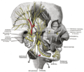
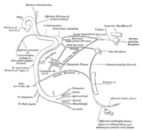
Plan of the facial and intermediate nerves and their communication with other nerves.
-

The course and connections of the facial nerve in the temporal bone.
-

Upper part of medulla spinalis and hind- and mid-brains; posterior aspect, exposed in situ.
-

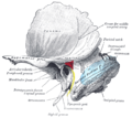
Left temporal bone showing surface markings for the tympanic antrum (red), transverse sinus (blue), and facial nerve (yellow).
-

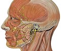
Head facial nerve branches
-

Facial nerve. Deep dissection.


See also[edit]
- List of medical mnemonics#Anatomy
- List of anatomy mnemonics#Cranial nerves
References[edit]
![]() This article incorporates text in the public domain from page 901 of the 20th edition of Gray’s Anatomy (1918)
This article incorporates text in the public domain from page 901 of the 20th edition of Gray’s Anatomy (1918)
- ^ a b «The Facial Nerve (CN VII)». TeachMeAnatomy. 2013-09-04. Retrieved 2018-05-04.
- ^ «Facial nerve | anatomy». Encyclopedia Britannica. Retrieved 2020-10-13.
- ^ Kochhar A, Larian B, Azizzadeh B (April 2016). «Facial Nerve and Parotid Gland Anatomy». Otolaryngologic Clinics of North America. 49 (2): 273–84. doi:10.1016/j.otc.2015.10.002. PMID 27040583.
- ^ Gupta S, Mends F, Hagiwara M, Fatterpekar G, Roehm PC (2013). «Imaging the facial nerve: a contemporary review». Radiology Research and Practice. 2013: 248039. doi:10.1155/2013/248039. PMC 3676972. PMID 23766904.
- ^ Snell RS (2011). Clinical Anatomy by Regions (Ninth ed.). Philadelphia, Pa.; London: LWW. ISBN 9781451110326.
- ^ Singh V. Textbook of Clinical Neuroanatomy (2nd ed.). p. 104.
- ^ Saha S, Pal S, Sengupta M, Chowdhury K, Saha VP, Mondal L (January 2014). «Identification of facial nerve during parotidectomy: a combined anatomical & surgical study». Indian Journal of Otolaryngology and Head and Neck Surgery. 66 (1): 63–8. doi:10.1007/s12070-013-0669-z. PMC 3938689. PMID 24605304.
- ^ Dudek RW (2014). BRS Embryology (Sixth ed.). LWW. ISBN 9781451190380.
- ^ Moore KL, Persaud TV, Torchia MG (2011). The Developing Human: Clinically Oriented Embryology with Student Consult Online Access, 9th Edition (9th ed.). Philadelphia, PA: Saunders. ISBN 9781437720020.
- ^ Masterson L, Vallis M, Quinlivan R, Prinsley P (September 2015). «Assessment and management of facial nerve palsy». BMJ. 351: h3725. doi:10.1136/bmj.h3725. PMID 26378061. S2CID 206906594.
- ^ Kliniska Färdigheter: Informationsutbytet Mellan Patient Och Läkare, LINDGREN, STEFAN, ISBN 91-44-37271-X
External links[edit]
From Wikipedia, the free encyclopedia
Not to be confused with other nerves that innervate the face, such as the trigeminal nerve.
| Facial nerve | |
|---|---|
|
The course of the facial nerve is shown here |
|
|
The nerves of the scalp, face, and side of neck. |
|
| Details | |
| From | facial nerve nucleus, intermediate nerve |
| To | greater superficial petrosal nerve, |
| Innervates | Motor: Muscles of facial expression, posterior belly of digastric, stylohyoid, stapedius Special sensory: taste to anterior two-thirds of tongue Parasympathetic: submandibular gland, sublingual gland, lacrimal glands |
| Identifiers | |
| Latin | nervus facialis |
| MeSH | D005154 |
| NeuroNames | 551 |
| TA98 | A14.2.01.099 |
| TA2 | 6284 |
| FMA | 50868 |
| Anatomical terms of neuroanatomy
[edit on Wikidata] |
The facial nerve, also known as the seventh cranial nerve, cranial nerve VII, or simply CN VII, is a cranial nerve that emerges from the pons of the brainstem, controls the muscles of facial expression, and functions in the conveyance of taste sensations from the anterior two-thirds of the tongue.[1][2] The nerve typically travels from the pons through the facial canal in the temporal bone and exits the skull at the stylomastoid foramen.[3] It arises from the brainstem from an area posterior to the cranial nerve VI (abducens nerve) and anterior to cranial nerve VIII (vestibulocochlear nerve).
The facial nerve also supplies preganglionic parasympathetic fibers to several head and neck ganglia.
The facial and intermediate nerves can be collectively referred to as the nervus intermediofacialis.
Structure[edit]
The path of the facial nerve can be divided into six segments:
- intracranial (cisternal) segment
- meatal (canalicular) segment (within the internal auditory canal)
- labyrinthine segment (internal auditory canal to geniculate ganglion)
- tympanic segment (from geniculate ganglion to pyramidal eminence)
- mastoid segment (from pyramidal eminence to stylomastoid foramen)
- extratemporal segment (from stylomastoid foramen to post parotid branches)
The motor part of the facial nerve arises from the facial nerve nucleus in the pons, while the sensory and parasympathetic parts of the facial nerve arise from the intermediate nerve.
From the brain stem, the motor and sensory parts of the facial nerve join together and traverse the posterior cranial fossa before entering the petrous temporal bone via the internal auditory meatus. Upon exiting the internal auditory meatus, the nerve then runs a tortuous course through the facial canal, which is divided into the labyrinthine, tympanic, and mastoid segments.
The labyrinthine segment is very short, and ends where the facial nerve forms a bend known as the geniculum of the facial nerve (genu meaning knee), which contains the geniculate ganglion for sensory nerve bodies. The first branch of the facial nerve, the greater petrosal nerve, arises here from the geniculate ganglion. The greater petrosal nerve runs through the pterygoid canal and synapses at the pterygopalatine ganglion. Postsynaptic fibers of the greater petrosal nerve innervate the lacrimal gland.
In the tympanic segment, the facial nerve runs through the tympanic cavity, medial to the incus.
The pyramidal eminence is the second bend in the facial nerve, where the nerve runs downward as the mastoid segment. In the temporal part of the facial canal, the nerve gives rise to the nerve to the stapedius muscle and chorda tympani. The chorda tympani supplies taste fibers to the anterior two thirds of the tongue, and also synapses with the submandibular ganglion. Postsynaptic fibers from the submandibular ganglion supply the sublingual and submandibular glands.
Upon emerging from the stylomastoid foramen, the facial nerve gives rise to the posterior auricular branch. The facial nerve then passes through the parotid gland, which it does not innervate, to form the parotid plexus, which splits into five branches (temporal, zygomatic, buccal, marginal mandibular, and cervical) innervating the muscles of facial expression.[4][5]
Intracranial branches[edit]
The greater petrosal nerve arises at the superior salivatory nucleus of the pons and provides parasympathetic innervation to several glands, including the nasal glands, the palatine glands, the lacrimal gland, and the pharyngeal gland. It also provides parasympathetic innervation to the sphenoid sinus, frontal sinus, maxillary sinus, ethmoid sinus, and nasal cavity. This nerve also includes taste fibers for the palate via the lesser palatine nerve and greater palatine nerve.
The communicating branch to the otic ganglion arises at the geniculate ganglion and joins the lesser petrosal nerve to reach the otic ganglion.[6]
The nerve to stapedius provides motor innervation for the stapedius muscle in middle ear
The chorda tympani provides parasympathetic innervation to the sublingual and submandibular glands, as well as special sensory taste fibers for the anterior two thirds of the tongue.[1]
[edit]
Distal to stylomastoid foramen, the following nerves branch off the facial nerve:
- Posterior auricular nerve which controls movements of some of the scalp muscles around the ear
- Branch to posterior belly of digastric muscle as well as the stylohyoid muscle
- Five major facial branches (at parotid plexus) – from superior to inferior:
- Temporal branch
- Zygomatic branch
- Buccal branch
- Marginal mandibular branch
- Cervical branch
Intra operatively the facial nerve is recognized at 3 constant landmarks:[citation needed]
- At the tip of tragus where the nerve is 1 cm deep and inferior[7]
- At the posterior belly of digastric by tracing this backwards to the tympanic plate, the nerve can be found between these two structures
- By locating the posterior facial vein at the inferior aspect of the gland where the marginal branch would be seen crossing it.
- Lateral semicircular canal
- Foot of incus
Nucleus[edit]
The cell bodies for the facial nerve are grouped in anatomical areas called nuclei or ganglia. The cell bodies for the afferent nerves are found in the geniculate ganglion for taste sensation. The cell bodies for muscular efferent nerves are found in the facial motor nucleus whereas the cell bodies for the parasympathetic efferent nerves are found in the superior salivatory nucleus.
Development[edit]
The facial nerve is developmentally derived from the second pharyngeal arch, or branchial arch. The second arch is called the hyoid arch because it contributes to the formation of the lesser horn and upper body of the hyoid bone (the rest of the hyoid is formed by the third arch). The facial nerve supplies motor and sensory innervation to the muscles formed by the second pharyngeal arch, including the muscles of facial expression, the posterior belly of the digastric, stylohyoid, and stapedius. The motor division of the facial nerve is derived from the basal plate of the embryonic pons, while the sensory division originates from the cranial neural crest.[8]
Although the anterior two thirds of the tongue are derived from the first pharyngeal arch, which gives rise to the trigeminal nerve, not all innervation of the tongue is supplied by it. The lingual branch of the mandibular division (V3) of the trigeminal nerve supplies non-taste sensation (pressure, heat, texture) to the anterior part of the tongue via general somatic afferent fibers. Nerve fibers for taste are supplied by the chorda tympani branch of the facial nerve via special visceral afferent fibers.[9]
Function[edit]
Facial expression[edit]
The main function of the facial nerve is motor control of all of the muscles of facial expression. It also innervates the posterior belly of the digastric muscle, the stylohyoid muscle, and the stapedius muscle of the middle ear. All of these muscles are striated muscles of branchiomeric origin developing from the 2nd pharyngeal arch.
Facial sensation[edit]
In addition, the facial nerve receives taste sensations from the anterior two-thirds of the tongue via the chorda tympani. Taste sensation is sent to the gustatory portion (superior part) of the solitary nucleus. General sensation from the anterior two-thirds of tongue are supplied by afferent fibers of the third division of the fifth cranial nerve (V-3). These sensory (V-3) and taste (VII) fibers travel together as the lingual nerve briefly before the chorda tympani leaves the lingual nerve to enter the tympanic cavity (middle ear) via the petrotympanic fissure. It joins the rest of the facial nerve via the canaliculus for chorda tympani. The facial nerve then forms the geniculate ganglion, which contains the cell bodies of the taste fibers of chorda tympani and other taste and sensory pathways. From the geniculate ganglion, the taste fibers continue as the intermediate nerve which goes to the upper anterior quadrant of the fundus of the internal acoustic meatus along with the motor root of the facial nerve. The intermediate nerve reaches the posterior cranial fossa via the internal acoustic meatus before synapsing in the solitary nucleus.
The facial nerve also supplies a small amount of afferent innervation to the oropharynx below the palatine tonsil. There is also a small amount of cutaneous sensation carried by the nervus intermedius from the skin in and around the auricle (outer ear).
Other[edit]
The facial nerve also supplies parasympathetic fibers to the submandibular gland and sublingual glands via chorda tympani. Parasympathetic innervation serves to increase the flow of saliva from these glands. It also supplies parasympathetic innervation to the nasal mucosa and the lacrimal gland via the pterygopalatine ganglion. The parasympathetic fibers that travel in the facial nerve originate in the superior salivatory nucleus.
The facial nerve also functions as the efferent limb of the corneal reflex.
Functional components[edit]
The facial nerve carries axons of type GSA, general somatic afferent, to skin of the posterior ear.
The facial nerve also carries axons of type GVE, general visceral efferent, which innervate the sublingual, submandibular, and lacrimal glands, also mucosa of nasal cavity.
Axons of type SVE, special visceral efferent, innervate muscles of facial expression, stapedius, the posterior belly of digastric, and the stylohyoid.
The axons of type SVA, special visceral afferent, provide taste to the anterior two-thirds of tongue via chorda tympani.
Clinical significance[edit]
Palsy[edit]
People may suffer from acute facial nerve paralysis, which is usually manifested by facial paralysis.[10] Bell’s palsy is one type of idiopathic acute facial nerve paralysis, which is more accurately described as a multiple cranial nerve ganglionitis that involves the facial nerve, and most likely results from viral infection and also sometimes as a result of Lyme disease. Iatrogenic Bell’s palsy may also be as a result of an incorrectly placed dental local-anesthetic (inferior alveolar nerve block). Although giving the appearance of a hemiplegic stroke, effects dissipate with the drug. When the facial nerve is permanently damaged due to a birth defect, trauma, or other disorder, surgery including a cross facial nerve graft or masseteric facial nerve transfer may be performed to help regain facial movement.[citation needed] Facial nerve decompression surgery is also sometimes carried out in certain cases of facial nerve compression.
Examination[edit]
Voluntary facial movements, such as wrinkling the brow, showing teeth, frowning, closing the eyes tightly (inability to do so is called lagophthalmos),[11] pursing the lips and puffing out the cheeks, all test the facial nerve. There should be no noticeable asymmetry.
In an upper motor neuron lesion, called central seven, only the lower part of the face on the contralateral side will be affected, due to the bilateral control to the upper facial muscles (frontalis and orbicularis oculi).
Lower motor neuron lesions can result in a CN VII palsy (Bell’s palsy is the idiopathic form of facial nerve palsy), manifested as both upper and lower facial weakness on the same side of the lesion.
Taste can be tested on the anterior two-thirds of the tongue. This can be tested with a swab dipped in a flavoured solution, or with electronic stimulation (similar to putting your tongue on a battery).
Corneal reflex. The afferent arc is mediated by the general sensory afferents of the trigeminal nerve. The efferent arc occurs via the facial nerve. The reflex involves consensual blinking of both eyes in response to stimulation of one eye. This is due to the facial nerves’ innervation of the muscles of facial expression, namely orbicularis oculi, responsible for blinking. Thus, the corneal reflex effectively tests the proper functioning of both cranial nerves V and VII.
Additional images[edit]
-
Inferior view of the human brain, with the cranial nerves labelled.
-
Mandibular division of the trifacial nerve.
-
Plan of the facial and intermediate nerves and their communication with other nerves.
-
The course and connections of the facial nerve in the temporal bone.
-
Upper part of medulla spinalis and hind- and mid-brains; posterior aspect, exposed in situ.
-
Left temporal bone showing surface markings for the tympanic antrum (red), transverse sinus (blue), and facial nerve (yellow).
-
Head facial nerve branches
-
Facial nerve. Deep dissection.
See also[edit]
- List of medical mnemonics#Anatomy
- List of anatomy mnemonics#Cranial nerves
References[edit]
![]() This article incorporates text in the public domain from page 901 of the 20th edition of Gray’s Anatomy (1918)
This article incorporates text in the public domain from page 901 of the 20th edition of Gray’s Anatomy (1918)
- ^ a b «The Facial Nerve (CN VII)». TeachMeAnatomy. 2013-09-04. Retrieved 2018-05-04.
- ^ «Facial nerve | anatomy». Encyclopedia Britannica. Retrieved 2020-10-13.
- ^ Kochhar A, Larian B, Azizzadeh B (April 2016). «Facial Nerve and Parotid Gland Anatomy». Otolaryngologic Clinics of North America. 49 (2): 273–84. doi:10.1016/j.otc.2015.10.002. PMID 27040583.
- ^ Gupta S, Mends F, Hagiwara M, Fatterpekar G, Roehm PC (2013). «Imaging the facial nerve: a contemporary review». Radiology Research and Practice. 2013: 248039. doi:10.1155/2013/248039. PMC 3676972. PMID 23766904.
- ^ Snell RS (2011). Clinical Anatomy by Regions (Ninth ed.). Philadelphia, Pa.; London: LWW. ISBN 9781451110326.
- ^ Singh V. Textbook of Clinical Neuroanatomy (2nd ed.). p. 104.
- ^ Saha S, Pal S, Sengupta M, Chowdhury K, Saha VP, Mondal L (January 2014). «Identification of facial nerve during parotidectomy: a combined anatomical & surgical study». Indian Journal of Otolaryngology and Head and Neck Surgery. 66 (1): 63–8. doi:10.1007/s12070-013-0669-z. PMC 3938689. PMID 24605304.
- ^ Dudek RW (2014). BRS Embryology (Sixth ed.). LWW. ISBN 9781451190380.
- ^ Moore KL, Persaud TV, Torchia MG (2011). The Developing Human: Clinically Oriented Embryology with Student Consult Online Access, 9th Edition (9th ed.). Philadelphia, PA: Saunders. ISBN 9781437720020.
- ^ Masterson L, Vallis M, Quinlivan R, Prinsley P (September 2015). «Assessment and management of facial nerve palsy». BMJ. 351: h3725. doi:10.1136/bmj.h3725. PMID 26378061. S2CID 206906594.
- ^ Kliniska Färdigheter: Informationsutbytet Mellan Patient Och Läkare, LINDGREN, STEFAN, ISBN 91-44-37271-X
External links[edit]
| В Википедии есть статья «лицевой нерв». |
Содержание
- 1 Русский
- 1.1 Тип и синтаксические свойства сочетания
- 1.2 Произношение
- 1.3 Семантические свойства
- 1.3.1 Значение
- 1.3.2 Синонимы
- 1.3.3 Антонимы
- 1.3.4 Гиперонимы
- 1.3.5 Гипонимы
- 1.4 Этимология
- 1.5 Перевод
- 1.6 Библиография
Русский[править]
Тип и синтаксические свойства сочетания[править]
ли—це—во́й нерв
Устойчивое сочетание (термин). Используется в качестве именной группы.
Произношение[править]
- МФА: [lʲɪt͡sɨˈvoɪ̯ nʲerf]
(файл)
Семантические свойства[править]
Значение[править]
- анат. седьмой (VII) из двенадцати черепных нервов, выходит из мозга между варолиевым мостом и продолговатым мозгом ◆ Отсутствует пример употребления (см. рекомендации).
Синонимы[править]
Антонимы[править]
Гиперонимы[править]
Гипонимы[править]
Этимология[править]
Перевод[править]
| Список переводов | |
Библиография[править]
Значение слова «ЛИЦЕВОЙ НЕРВ» найдено в 18 источниках
ЛИЦЕВОЙ НЕРВ
(Nervus facialis) — см. Личной нерв.
VII пара черепно-мозговых нервов, смешанный нерв, содержащий двигательные и чувствительные нервные волокна. Ядра Л. н. заложены в варолиевом мосту (см. рис. 1 в ст. Головной мозг,); проводники, связанные с этими ядрами, формируют ствол Л. н., который, пройдя через внутренний слуховой проход и пирамиду височной кости, выходит из полости черепа через шило-сосцевидное отверстие. В веществе околоушной слюнной железы он делится на свои конечные ветви. Двигательные проводники Л. н. иннервируют мимическую мускулатуру, шило-подъязычную мышцу, заднее брюшко двубрюшной мышцы и подкожную мышцу шеи. Вегетативные волокна Л. н. иннервируют слюнные железы, слёзную железу, железы слизистой оболочки полости носа, нёба и верхнего отдела глотки. Чувствительные волокна Л. н. обеспечивают вкусовую иннервацию передних двух третей языка. Вегетативные и чувствительные волокна Л. н. вблизи от ствола мозга формируют особый промежуточный нерв, наиболее крупная ветвь которого — т. н. барабанная струна. Наиболее частая патология — паралич.
Лицевой нерв (Nervus facialis) — см. Личной нерв.
ЛИЦЕВОЙ НЕРВ
(nervus facialis), VII пара черепномозговых нервов; смешанный нерв.
.(Источник: «Биологический энциклопедический словарь.» Гл. ред. М. С. Гиляров; Редкол.: А. А. Бабаев, Г. Г. Винберг, Г. А. Заварзин и др. — 2-е изд., исправл. — М.: Сов. Энциклопедия, 1986.)
ЛИЦЕВОЙ НЕРВ, седьмая пара черепных нервов у позвоночных животных и человека; содержит двигательные и чувствительные нервные волокна. У человека иннервирует мимическую мускулатуру, слезные и слюнные железы, слизистую оболочку языка, неба, полости носа и верхнего отдела глотки.
ЛИЦЕВОЙ НЕРВ — седьмая пара черепных нервов у позвоночных животных и человека; содержит двигательные и чувствительные нервные волокна. У человека иннервирует мимическую мускулатуру, слезные и слюнные железы, слизистую оболочку языка, неба, полости носа и верхнего отдела глотки.
ЛИЦЕВОЙ НЕРВ , седьмая пара черепных нервов у позвоночных животных и человека; содержит двигательные и чувствительные нервные волокна. У человека иннервирует мимическую мускулатуру, слезные и слюнные железы, слизистую оболочку языка, неба, полости носа и верхнего отдела глотки.
ЛИЦЕВОЙ НЕРВ, седьмая пара черепных нервов у позвоночных животных и человека; содержит двигательные и чувствительные нервные волокна. У человека иннервирует мимическую мускулатуру, слезные и слюнные железы, слизистую оболочку языка, неба, полости носа и верхнего отдела глотки.
— седьмая пара черепных нервов у позвоночных животных ичеловека; содержит двигательные и чувствительные нервные волокна. Учеловека иннервирует мимическую мускулатуру, слезные и слюнные железы,слизистую оболочку языка, неба, полости носа и верхнего отдела глотки.
седьмая пара черепных нервов у позвоночных животных и человека; содержит двигат. и чувствит. нерв. волокна. У человека ипвервирует мимич. мускулатуру, слёзные и слюнные железы, слизистую оболочку языка, нёба, полости носа и верх. отдела глотки.
VII черепной нерв. Смешанный нерв с эфферентными волокнами, идущими к мимическим мышцам, подкожной мышце шеи и подъязычным железам, и с афферентными волокнами, идущими от вкусовых сосочков передних двух третей языка.
7 черепной нерв смешанного типа. Афферентные его волокна иннервируют мимические мышцы, подкожную мышцу шеи и подъязычные железы, эфферентные волокна идут от вкусовых сосочков передних двух третей языка.
facial [VII cranial] nerve, nervus facialis [NA]
facial nerve, VII cranial nerve
1) nerf crânien septième
2) nerf facial
(nervus facialis) nervo facciale
- Подбор слов
- Значения слов
- лицевой нерв
Поиск значения / толкования слов
Раздел очень прост в использовании. В предложенное поле достаточно ввести нужное слово, и мы вам выдадим
список его значений. Хочется отметить, что
наш сайт предоставляет данные из разных источников – энциклопедического, толкового, словообразовательного
словарей. Также здесь можно познакомиться
с примерами употребления введенного вами слова.
Энциклопедический словарь, 1998 г.
лицевой нерв
седьмая пара черепных нервов у позвоночных животных и человека; содержит двигательные и чувствительные нервные волокна. У человека иннервирует мимическую мускулатуру, слезные и слюнные железы, слизистую оболочку языка, неба, полости носа и верхнего отдела глотки.
Большая Советская Энциклопедия
Лицевой нерв
(Nervus facialis), VII пара черепно-мозговых нервов, смешанный нерв, содержащий двигательные и чувствительные нервные волокна. Ядра Л. н. заложены в варолиевом мосту (см. рис. 1 в ст. Головной мозг ,); проводники, связанные с этими ядрами, формируют ствол Л. н., который, пройдя через внутренний слуховой проход и пирамиду височной кости, выходит из полости черепа через шило-сосцевидное отверстие. В веществе околоушной слюнной железы он делится на свои конечные ветви. Двигательные проводники Л. н. иннервируют мимическую мускулатуру, шило-подъязычную мышцу, заднее брюшко двубрюшной мышцы и подкожную мышцу шеи. Вегетативные волокна Л. н. иннервируют слюнные железы, слёзную железу, железы слизистой оболочки полости носа, нёба и верхнего отдела глотки. Чувствительные волокна Л. н. обеспечивают вкусовую иннервацию передних двух третей языка. Вегетативные и чувствительные волокна Л. н. вблизи от ствола мозга формируют особый промежуточный нерв, наиболее крупная ветвь которого ≈ т. н. барабанная струна. Наиболее частая патология ≈ паралич.
Википедия
Лицевой нерв
Лицевой нерв , седьмой (VII) из двенадцати черепных нервов , выходит из мозга между варолиевым мостом и продолговатым мозгом . Лицевой нерв иннервирует мимические мышцы лица. Также в составе лицевого нерва проходит промежуточный нерв ответственный за иннервацию слёзной железы, стременной мышцы и вкусовой чувствительности двух передних третей языка .