From Wikipedia, the free encyclopedia
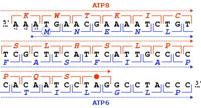
Stop codon (red dot) of the human mitochondrial DNA MT-ATP8 gene, and start codon (blue circle) of the MT-ATP6 gene. For each nucleotide triplet (square brackets), the corresponding amino acid is given (one-letter code), either in the +1 reading frame for MT-ATP8 (in red) or in the +3 frame for MT-ATP6 (in blue). In this genomic region, the two genes overlap.
In molecular biology (specifically protein biosynthesis), a stop codon (or termination codon) is a codon (nucleotide triplet within messenger RNA) that signals the termination of the translation process of the current protein.[1] Most codons in messenger RNA correspond to the addition of an amino acid to a growing polypeptide chain, which may ultimately become a protein; stop codons signal the termination of this process by binding release factors, which cause the ribosomal subunits to disassociate, releasing the amino acid chain.
While start codons need nearby sequences or initiation factors to start translation, a stop codon alone is sufficient to initiate termination.
Properties[edit]
Standard codons[edit]
In the standard genetic code, there are three different termination codons:
| Codon | Standard code (Translation table 1) |
Name | ||
|---|---|---|---|---|
| DNA | RNA | |||
TAG |
UAG
|
STOP = Ter (*)
|
«amber» | |
TAA |
UAA
|
STOP = Ter (*)
|
«ochre» | |
TGA |
UGA
|
STOP = Ter (*)
|
«opal» (or «umber») |
Alternative stop codons[edit]
There are variations on the standard genetic code, and alternative stop codons have been found in the mitochondrial genomes of vertebrates,[2] Scenedesmus obliquus,[3] and Thraustochytrium.[4]
| Genetic code | Translation table |
Codon | Translation with this code |
Standard translation | ||
|---|---|---|---|---|---|---|
| DNA | RNA | |||||
| Vertebrate mitochondrial | 2 | AGA |
AGA |
STOP = Ter (*) |
Arg (R)
|
|
AGG |
AGG |
STOP = Ter (*) |
Arg (R)
|
|||
| Scenedesmus obliquus mitochondrial | 22 | TCA |
UCA |
STOP = Ter (*) |
Ser (S)
|
|
| Thraustochytrium mitochondrial | 23 | TTA |
UUA |
STOP = Ter (*) |
Leu (L)
|
| Amino-acid biochemical properties | Nonpolar | Polar | Basic | Acidic | Termination: stop codon |
Reassigned stop codons[edit]
The nuclear genetic code is flexible as illustrated by variant genetic codes that reassign standard stop codons to amino acids.[5]
| Genetic code | Translation table |
Codon | Conditional translation |
Standard translation | ||||
|---|---|---|---|---|---|---|---|---|
| DNA | RNA | |||||||
| Karyorelict nuclear | 27 | TGA |
UGA |
Ter (*) |
or | Trp (W) |
Ter (*)
|
|
| Condylostoma nuclear | 28 | TAA |
UAA |
Ter (*) |
or | Gln (Q) |
Ter (*)
|
|
TAG |
UAG |
Ter (*) |
or | Gln (Q) |
Ter (*)
|
|||
TGA |
UGA |
Ter (*) |
or | Trp (W) |
Ter (*)
|
|||
| Blastocrithidia nuclear | 31 | TAA |
UAA |
Ter (*) |
or | Glu (E) |
Ter (*)
|
|
TAG |
UAG |
Ter (*) |
or | Glu (E) |
Ter (*)
|
Translation[edit]
In 1986, convincing evidence was provided that selenocysteine (Sec) was incorporated co-translationally. Moreover, the codon partially directing its incorporation in the polypeptide chain was identified as UGA also known as the opal termination codon.[6] Different mechanisms for overriding the termination function of this codon have been identified in prokaryotes and in eukaryotes.[7] A particular difference between these kingdoms is that cis elements seem restricted to the neighborhood of the UAG codon in prokaryotes while in eukaryotes this restriction is not present. Instead such locations seem disfavored albeit not prohibited. [8]
In 2003, a landmark paper described the identification of all known selenoproteins in humans: 25 in total.[9] Similar analyses have been run for other organisms.
The UAG codon can translate into pyrrolysine (Pyl) in a similar manner.
Genomic distribution[edit]
Distribution of stop codons within the genome of an organism is non-random and can correlate with GC-content.[10][11] For example, the E. coli K-12 genome contains 2705 TAA (63%), 1257 TGA (29%), and 326 TAG (8%) stop codons (GC content 50.8%).[12] Also the substrates for the stop codons release factor 1 or release factor 2 are strongly correlated to the abundance of stop codons.[11] Large scale study of bacteria with a broad range of GC-contents shows that while the frequency of occurrence of TAA is negatively correlated to the GC-content and the frequency of occurrence of TGA is positively correlated to the GC-content, the frequency of occurrence of the TAG stop codon, which is often the minimally used stop codon in a genome, is not influenced by the GC-content.[13]
Recognition[edit]
Recognition of stop codons in bacteria have been associated with the so-called ‘tripeptide anticodon’,[14] a highly conserved amino acid motif in RF1 (PxT) and RF2 (SPF). Even though this is supported by structural studies, it was shown that the tripeptide anticodon hypothesis is an oversimplification.[15]
Nomenclature[edit]
Stop codons were historically given many different names, as they each corresponded to a distinct class of mutants that all behaved in a similar manner. These mutants were first isolated within bacteriophages (T4 and lambda), viruses that infect the bacteria Escherichia coli. Mutations in viral genes weakened their infectious ability, sometimes creating viruses that were able to infect and grow within only certain varieties of E. coli.
amber mutations (UAG)[edit]
They were the first set of nonsense mutations to be discovered, isolated by Richard H. Epstein and Charles Steinberg and named after their friend and graduate Caltech student Harris Bernstein, whose last name means «amber» in German (cf. Bernstein).[16][17]
Viruses with amber mutations are characterized by their ability to infect only certain strains of bacteria, known as amber suppressors. These bacteria carry their own mutation that allows a recovery of function in the mutant viruses. For example, a mutation in the tRNA that recognizes the amber stop codon allows translation to «read through» the codon and produce a full-length protein, thereby recovering the normal form of the protein and «suppressing» the amber mutation.[18]
Thus, amber mutants are an entire class of virus mutants that can grow in bacteria that contain amber suppressor mutations. Similar suppressors are known for ochre and opal stop codons as well.
tRNA molecules carrying unnatural aminoacids have been designed to recognize the amber stop codon in bacterial RNA. This technology allows for incorporation of orthogonal aminoacids (such as p-azidophenylalanine) at specific locations of the target protein.
ochre mutations (UAA)[edit]
It was the second stop codon mutation to be discovered. Reminiscent of the usual yellow-orange-brown color associated with amber, this second stop codon was given the name of «ochre», an orange-reddish-brown mineral pigment.[17]
Ochre mutant viruses had a property similar to amber mutants in that they recovered infectious ability within certain suppressor strains of bacteria. The set of ochre suppressors was distinct from amber suppressors, so ochre mutants were inferred to correspond to a different nucleotide triplet. Through a series of mutation experiments comparing these mutants with each other and other known amino acid codons, Sydney Brenner concluded that the amber and ochre mutations corresponded to the nucleotide triplets «UAG» and «UAA».[19]
opal or umber mutations (UGA)[edit]
The third and last stop codon in the standard genetic code was discovered soon after, and corresponds to the nucleotide triplet «UGA».[20]
To continue matching with the theme of colored minerals, the third nonsense codon came to be known as «opal», which is a type of silica showing a variety of colors.[17] Nonsense mutations that created this premature stop codon were later called opal mutations or umber mutations.
Mutations and disease[edit]
Nonsense[edit]
Nonsense mutations are changes in DNA sequence that introduce a premature stop codon, causing any resulting protein to be abnormally shortened. This often causes a loss of function in the protein, as critical parts of the amino acid chain are no longer assembled. Because of this terminology, stop codons have also been referred to as nonsense codons.
Nonstop[edit]
A nonstop mutation, also called a stop-loss variant, is a point mutation that occurs within a stop codon. Nonstop mutations cause the continued translation of an mRNA strand into what should be an untranslated region. Most polypeptides resulting from a gene with a nonstop mutation lose their function due to their extreme length and the impact on normal folding. Nonstop mutations differ from nonsense mutations in that they do not create a stop codon but, instead, delete one. Nonstop mutations also differ from missense mutations, which are point mutations where a single nucleotide is changed to cause replacement by a different amino acid. Nonstop mutations have been linked with many inherited diseases including endocrine disorders,[21] eye disease,[22] and neurodevelopmental disorders.[23][24]
Hidden stops[edit]
An example of a single base deletion forming a stop codon.
Hidden stops are non-stop codons that would be read as stop codons if they were frameshifted +1 or −1. These prematurely terminate translation if the corresponding frame-shift (such as due to a ribosomal RNA slip) occurs before the hidden stop. It is hypothesised that this decreases resource wastage on nonfunctional proteins and the production of potential cytotoxins. Researchers at Louisiana State University propose the ambush hypothesis, that hidden stops are selected for. Codons that can form hidden stops are used in genomes more frequently compared to synonymous codons that would otherwise code for the same amino acid. Unstable rRNA in an organism correlates with a higher frequency of hidden stops.[25]
However, this hypothesis could not be validated with a larger data set.[26]
Stop-codons and hidden stops together are collectively referred as stop-signals. Researchers at University of Memphis found that the ratios of the stop-signals on the three reading frames of a genome (referred to as translation stop-signals ratio or TSSR) of genetically related bacteria, despite their great differences in gene contents, are much alike. This nearly identical genomic-TSSR value of genetically related bacteria may suggest that bacterial genome expansion is limited by their unique stop-signals bias of that bacterial species.[27]
Translational readthrough[edit]
Stop codon suppression or translational readthrough occurs when in translation a stop codon is interpreted as a sense codon, that is, when a (standard) amino acid is ‘encoded’ by the stop codon. Mutated tRNAs can be the cause of readthrough, but also certain nucleotide motifs close to the stop codon. Translational readthrough is very common in viruses and bacteria, and has also been found as a gene regulatory principle in humans, yeasts, bacteria and drosophila.[28][29] This kind of endogenous translational readthrough constitutes a variation of the genetic code, because a stop codon codes for an amino acid. In the case of human malate dehydrogenase, the stop codon is read through with a frequency of about 4%.[30] The amino acid inserted at the stop codon depends on the identity of the stop codon itself: Gln, Tyr, and Lys have been found for the UAA and UAG codons, while Cys, Trp, and Arg for the UGA codon have been identified by mass spectrometry.[31] Extent of readthrough in mammals have widely variable extents, and can broadly diversify the proteome and affect cancer progression.[32]
Use as a watermark[edit]
In 2010 when Craig Venter unveiled the first fully functioning, reproducing cell controlled by synthetic DNA he described how his team used frequent stop codons to create watermarks in RNA and DNA to help confirm the results were indeed synthetic (and not contaminated or otherwise), using it to encode authors’ names and website addresses.[33]
See also[edit]
- Genetic code
- Start codon
- Terminator gene
References[edit]
- ^ Griffiths AJF, Miller JH, Suzuki DT, Lewontin RC, Gelbart WM (2000). «Chapter 10 (Molecular Biology of Gene Function): Genetic code: Stop codons». An Introduction to Genetic Analysis. W.H. Freeman and Company.
- ^ Barrell, B. G.; Bankier, A. T.; Drouin, J. (1979-11-08). «A different genetic code in human mitochondria». Nature. 282 (5735): 189–194. Bibcode:1979Natur.282..189B. doi:10.1038/282189a0. ISSN 0028-0836. PMID 226894. S2CID 4335828.
- ^ A. M. Nedelcu, R. W. Lee, G. Lemieux, M. W. Gray, G. Burger (June 2000). «The complete mitochondrial DNA sequence of Scenedesmus obliquus reflects an intermediate stage in the evolution of the green algal mitochondrial genome». Genome Research. 10 (6): 819–831. doi:10.1101/gr.10.6.819. PMC 310893. PMID 10854413.
{{cite journal}}: CS1 maint: multiple names: authors list (link) - ^ Wideman, Jeremy G.; Monier, Adam; Rodríguez-Martínez, Raquel; Leonard, Guy; Cook, Emily; Poirier, Camille; Maguire, Finlay; Milner, David S.; Irwin, Nicholas A. T.; Moore, Karen; Santoro, Alyson E. (2019-11-25). «Unexpected mitochondrial genome diversity revealed by targeted single-cell genomics of heterotrophic flagellated protists». Nature Microbiology. 5 (1): 154–165. doi:10.1038/s41564-019-0605-4. hdl:10871/39819. ISSN 2058-5276. PMID 31768028. S2CID 208279678.
- ^ Swart, Estienne Carl; Serra, Valentina; Petroni, Giulio; Nowacki, Mariusz (2016). «Genetic Codes with No Dedicated Stop Codon: Context-Dependent Translation Termination». Cell. 166 (3): 691–702. doi:10.1016/j.cell.2016.06.020. PMC 4967479. PMID 27426948.
- ^ Zinoni, F; Birkmann, A; Stadtman, T; Böck, A (1986). «Nucleotide sequence and expression of the selenocysteine-containing polypeptide of formate dehydrogenase (formate-hydrogen-lyase-linked) from Escherichia coli». Proceedings of the National Academy of Sciences. 83 (13): 4650–4654. Bibcode:1986PNAS…83.4650Z. doi:10.1073/pnas.83.13.4650. PMC 323799. PMID 2941757.
- ^ Böck, A (2013). «Selenoprotein Synthesis». Encyclopedia of Biological Chemistry. pp. 210–213. doi:10.1016/B978-0-12-378630-2.00025-6. ISBN 9780123786319. Retrieved 23 August 2021.
- ^ Mix, H; Lobanov, A; Gladyshev, V (2007). «SECIS elements in the coding regions of selenoprotein transcripts are functional in higher eukaryotes». Nucleic Acids Research. 35 (2): 414–423. doi:10.1093/nar/gkl1060. PMC 1802603. PMID 17169995.
- ^ Kryukov, G; Gladyshev, V (2003). «Characterization of mammalian selenoproteomes». Science. 300 (5624): 1439–1443. Bibcode:2003Sci…300.1439K. doi:10.1126/science.1083516. PMID 12775843. S2CID 10363908.
- ^ Povolotskaya IS, Kondrashov FA, Ledda A, Vlasov PK (2012). «Stop codons in bacteria are not selectively equivalent». Biology Direct. 7: 30. doi:10.1186/1745-6150-7-30. PMC 3549826. PMID 22974057.
- ^ a b Korkmaz, Gürkan; Holm, Mikael; Wiens, Tobias; Sanyal, Suparna (2014). «Comprehensive Analysis of Stop Codon Usage in Bacteria and Its Correlation with Release Factor Abundance». The Journal of Biological Chemistry. 289 (44): 775–806. doi:10.1074/jbc.M114.606632. PMC 4215218. PMID 25217634.
- ^ «Escherichia coli str. K-12 substr. MG1655, complete genome [Genbank Accession Number: U00096]». GenBank. NCBI. Retrieved 2013-01-27.
- ^ Wong, Tit-Yee; Fernandes, Sanjit; Sankhon, Naby; Leong, Patrick P; Kuo, Jimmy; Liu, Jong-Kang (2008). «Role of Premature Stop Codons in Bacterial Evolution». Journal of Bacteriology. 190 (20): 6718–6725. doi:10.1128/JB.00682-08. PMC 2566208. PMID 18708500.
- ^ Ito, Koichi; Uno, Makiko; Nakamura, Yoshikazu (1999). «A tripeptide ‘anticodon’ deciphers stop codons in messenger RNA». Nature. 403 (6770): 680–684. doi:10.1038/35001115. PMID 10688208. S2CID 4331695.
- ^ Korkmaz, Gürkan; Sanyal, Suparna (2017). «R213I mutation in release factor 2 (RF2) is one step forward for engineering an omnipotent release factor in bacteria Escherichia coli«. Journal of Biological Chemistry. 292 (36): 15134–15142. doi:10.1074/jbc.M117.785238. PMC 5592688. PMID 28743745.
- ^ Stahl FW (1995). «The amber mutants of phage T4». Genetics. 141 (2): 439–442. doi:10.1093/genetics/141.2.439. PMC 1206745. PMID 8647382.
- ^ a b c Lewin, Benjamin; Krebs, Jocelyn E.; Goldstein, Elliott S.; Kilpatrick, Stephen T. (2011-04-18). Lewin’s Essential GENES. Jones & Bartlett Publishers. ISBN 978-1-4496-4380-5.
- ^ Robin Cook. «Amber, Ocher, and Opal Mutations Summary». World of Genetics. Gale.
- ^ Brenner, S.; Stretton, A. O. W.; Kaplan, S. (1965). «Genetic Code: The ‘Nonsense’ Triplets for Chain Termination and their Suppression». Nature. 206 (4988): 994–8. Bibcode:1965Natur.206..994B. doi:10.1038/206994a0. PMID 5320272. S2CID 28502898.
- ^ Brenner, S.; Barnett, L.; Katz, E. R.; Crick, F. H. C. (1967). «UGA: A Third Nonsense Triplet in the Genetic Code». Nature. 213 (5075): 449–50. Bibcode:1967Natur.213..449B. doi:10.1038/213449a0. PMID 6032223. S2CID 4211867.
- ^ Pang S.; Wang W.; et al. (2002). «A novel nonstop mutation in the stop codon and a novel missense mutation in the type II 3beta-hydroxysteroid dehydrogenase (3beta-HSD) gene causing, respectively, nonclassic and classic 3beta-HSD deficiency congenital adrenal hyperplasia». J Clin Endocrinol Metab. 87 (6): 2556–63. doi:10.1210/jcem.87.6.8559. PMID 12050213.
- ^ Doucette, L.; et al. (2011). «A novel, non-stop mutation in FOXE3 causes an autosomal dominant form of variable anterior segment dysgenesis including Peters anomaly». European Journal of Human Genetics. 19 (3): 293–299. doi:10.1038/ejhg.2010.210. PMC 3062009. PMID 21150893.
- ^ Torres-Torronteras, J.; Rodriguez-Palmero, A.; et al. (2011). «A novel nonstop mutation in TYMP does not induce nonstop mRNA decay in a MNGIE patient with severe neuropathy» (PDF). Hum. Mutat. 32 (4): E2061–E2068. doi:10.1002/humu.21447. PMID 21412940. S2CID 24446773.
- ^ Spaull, R; Steel, D; Barwick, K; Prabhakar, P; Wakeling, E; Kurian, MA (2022-07-23). «STXBP1 Stop‐Loss Mutation Associated with Complex Early Onset Movement Disorder without Epilepsy». Movement Disorders Clinical Practice. 9 (6): 837–840. doi:10.1002/mdc3.13509. ISSN 2330-1619. PMC 9346254. PMID 35937496.
- ^ Seligmann, Hervé; Pollock, David D. (2004). «The Ambush Hypothesis: Hidden Stop Codons Prevent Off-Frame Gene Reading». DNA and Cell Biology. 23 (10): 701–5. doi:10.1089/1044549042476910. PMID 15585128.
- ^ Cavalcanti, Andre; Chang, Charlotte H.; Morgens, David W. (2013). «Ambushing the ambush hypothesis: predicting and evaluating off-frame codon frequencies in Prokaryotic Genomes». BMC Genomics. 14 (418): 1–8. doi:10.1186/1471-2164-14-418. PMC 3700767. PMID 23799949.
- ^ Wong, Tit-Yee; Schwartzbach, Steve (2015). «Protein mis-termination initiates genetic diseases, cancers, and restricts bacterial genome expansion». Journal of Environmental Science and Health, Part C. 33 (3): 255–85. doi:10.1080/10590501.2015.1053461. PMID 26087060. S2CID 20380447.
- ^ Namy O, Rousset JP, Napthine S, Brierley I (2004). «Reprogrammed genetic decoding in cellular gene expression». Molecular Cell. 13 (2): 157–68. doi:10.1016/S1097-2765(04)00031-0. PMID 14759362.
- ^ Schueren F, Lingner T, George R, Hofhuis J, Gartner J, Thoms S (2014). «Peroxisomal lactate dehydrogenase is generated by translational readthrough in mammals». eLife. 3: e03640. doi:10.7554/eLife.03640. PMC 4359377. PMID 25247702.
- ^ Hofhuis J, Schueren F, Nötzel C, Lingner T, Gärtner J, Jahn O, Thoms S (2016). «The functional readthrough extension of malate dehydrogenase reveals a modification of the genetic code». Open Biol. 6 (11): 160246. doi:10.1098/rsob.160246. PMC 5133446. PMID 27881739.
- ^ Blanchet S, Cornu D, Argentini M, Namy O (2014). «New insights into the incorporation of natural suppressor tRNAs at stop codons in Saccharomyces cerevisiae«. Nucleic Acids Res. 42 (15): 10061–72. doi:10.1093/nar/gku663. PMC 4150775. PMID 25056309.
- ^ Ghosh, Souvik; Guimaraes, Joao C; Lanzafame, Manuela; Schmidt, Alexander; Syed, Afzal Pasha; Dimitriades, Beatrice; Börsch, Anastasiya; Ghosh, Shreemoyee; Mittal, Nitish; Montavon, Thomas; Correia, Ana Luisa; Danner, Johannes; Meister, Gunter; Terracciano, Luigi M; Pfeffer, Sébastien; Piscuoglio, Salvatore; Zavolan, Mihaela (15 September 2020). «Prevention of dsRNA‐induced interferon signaling by AGO1x is linked to breast cancer cell proliferation». The EMBO Journal. 39 (18): e103922. doi:10.15252/embj.2019103922. PMC 7507497. PMID 32812257.
- ^ «Watch me unveil «synthetic life»«. 21 May 2010.
From Wikipedia, the free encyclopedia
Stop codon (red dot) of the human mitochondrial DNA MT-ATP8 gene, and start codon (blue circle) of the MT-ATP6 gene. For each nucleotide triplet (square brackets), the corresponding amino acid is given (one-letter code), either in the +1 reading frame for MT-ATP8 (in red) or in the +3 frame for MT-ATP6 (in blue). In this genomic region, the two genes overlap.
In molecular biology (specifically protein biosynthesis), a stop codon (or termination codon) is a codon (nucleotide triplet within messenger RNA) that signals the termination of the translation process of the current protein.[1] Most codons in messenger RNA correspond to the addition of an amino acid to a growing polypeptide chain, which may ultimately become a protein; stop codons signal the termination of this process by binding release factors, which cause the ribosomal subunits to disassociate, releasing the amino acid chain.
While start codons need nearby sequences or initiation factors to start translation, a stop codon alone is sufficient to initiate termination.
Properties[edit]
Standard codons[edit]
In the standard genetic code, there are three different termination codons:
| Codon | Standard code (Translation table 1) |
Name | ||
|---|---|---|---|---|
| DNA | RNA | |||
TAG |
UAG
|
STOP = Ter (*)
|
«amber» | |
TAA |
UAA
|
STOP = Ter (*)
|
«ochre» | |
TGA |
UGA
|
STOP = Ter (*)
|
«opal» (or «umber») |
Alternative stop codons[edit]
There are variations on the standard genetic code, and alternative stop codons have been found in the mitochondrial genomes of vertebrates,[2] Scenedesmus obliquus,[3] and Thraustochytrium.[4]
| Genetic code | Translation table |
Codon | Translation with this code |
Standard translation | ||
|---|---|---|---|---|---|---|
| DNA | RNA | |||||
| Vertebrate mitochondrial | 2 | AGA |
AGA |
STOP = Ter (*) |
Arg (R)
|
|
AGG |
AGG |
STOP = Ter (*) |
Arg (R)
|
|||
| Scenedesmus obliquus mitochondrial | 22 | TCA |
UCA |
STOP = Ter (*) |
Ser (S)
|
|
| Thraustochytrium mitochondrial | 23 | TTA |
UUA |
STOP = Ter (*) |
Leu (L)
|
| Amino-acid biochemical properties | Nonpolar | Polar | Basic | Acidic | Termination: stop codon |
Reassigned stop codons[edit]
The nuclear genetic code is flexible as illustrated by variant genetic codes that reassign standard stop codons to amino acids.[5]
| Genetic code | Translation table |
Codon | Conditional translation |
Standard translation | ||||
|---|---|---|---|---|---|---|---|---|
| DNA | RNA | |||||||
| Karyorelict nuclear | 27 | TGA |
UGA |
Ter (*) |
or | Trp (W) |
Ter (*)
|
|
| Condylostoma nuclear | 28 | TAA |
UAA |
Ter (*) |
or | Gln (Q) |
Ter (*)
|
|
TAG |
UAG |
Ter (*) |
or | Gln (Q) |
Ter (*)
|
|||
TGA |
UGA |
Ter (*) |
or | Trp (W) |
Ter (*)
|
|||
| Blastocrithidia nuclear | 31 | TAA |
UAA |
Ter (*) |
or | Glu (E) |
Ter (*)
|
|
TAG |
UAG |
Ter (*) |
or | Glu (E) |
Ter (*)
|
Translation[edit]
In 1986, convincing evidence was provided that selenocysteine (Sec) was incorporated co-translationally. Moreover, the codon partially directing its incorporation in the polypeptide chain was identified as UGA also known as the opal termination codon.[6] Different mechanisms for overriding the termination function of this codon have been identified in prokaryotes and in eukaryotes.[7] A particular difference between these kingdoms is that cis elements seem restricted to the neighborhood of the UAG codon in prokaryotes while in eukaryotes this restriction is not present. Instead such locations seem disfavored albeit not prohibited. [8]
In 2003, a landmark paper described the identification of all known selenoproteins in humans: 25 in total.[9] Similar analyses have been run for other organisms.
The UAG codon can translate into pyrrolysine (Pyl) in a similar manner.
Genomic distribution[edit]
Distribution of stop codons within the genome of an organism is non-random and can correlate with GC-content.[10][11] For example, the E. coli K-12 genome contains 2705 TAA (63%), 1257 TGA (29%), and 326 TAG (8%) stop codons (GC content 50.8%).[12] Also the substrates for the stop codons release factor 1 or release factor 2 are strongly correlated to the abundance of stop codons.[11] Large scale study of bacteria with a broad range of GC-contents shows that while the frequency of occurrence of TAA is negatively correlated to the GC-content and the frequency of occurrence of TGA is positively correlated to the GC-content, the frequency of occurrence of the TAG stop codon, which is often the minimally used stop codon in a genome, is not influenced by the GC-content.[13]
Recognition[edit]
Recognition of stop codons in bacteria have been associated with the so-called ‘tripeptide anticodon’,[14] a highly conserved amino acid motif in RF1 (PxT) and RF2 (SPF). Even though this is supported by structural studies, it was shown that the tripeptide anticodon hypothesis is an oversimplification.[15]
Nomenclature[edit]
Stop codons were historically given many different names, as they each corresponded to a distinct class of mutants that all behaved in a similar manner. These mutants were first isolated within bacteriophages (T4 and lambda), viruses that infect the bacteria Escherichia coli. Mutations in viral genes weakened their infectious ability, sometimes creating viruses that were able to infect and grow within only certain varieties of E. coli.
amber mutations (UAG)[edit]
They were the first set of nonsense mutations to be discovered, isolated by Richard H. Epstein and Charles Steinberg and named after their friend and graduate Caltech student Harris Bernstein, whose last name means «amber» in German (cf. Bernstein).[16][17]
Viruses with amber mutations are characterized by their ability to infect only certain strains of bacteria, known as amber suppressors. These bacteria carry their own mutation that allows a recovery of function in the mutant viruses. For example, a mutation in the tRNA that recognizes the amber stop codon allows translation to «read through» the codon and produce a full-length protein, thereby recovering the normal form of the protein and «suppressing» the amber mutation.[18]
Thus, amber mutants are an entire class of virus mutants that can grow in bacteria that contain amber suppressor mutations. Similar suppressors are known for ochre and opal stop codons as well.
tRNA molecules carrying unnatural aminoacids have been designed to recognize the amber stop codon in bacterial RNA. This technology allows for incorporation of orthogonal aminoacids (such as p-azidophenylalanine) at specific locations of the target protein.
ochre mutations (UAA)[edit]
It was the second stop codon mutation to be discovered. Reminiscent of the usual yellow-orange-brown color associated with amber, this second stop codon was given the name of «ochre», an orange-reddish-brown mineral pigment.[17]
Ochre mutant viruses had a property similar to amber mutants in that they recovered infectious ability within certain suppressor strains of bacteria. The set of ochre suppressors was distinct from amber suppressors, so ochre mutants were inferred to correspond to a different nucleotide triplet. Through a series of mutation experiments comparing these mutants with each other and other known amino acid codons, Sydney Brenner concluded that the amber and ochre mutations corresponded to the nucleotide triplets «UAG» and «UAA».[19]
opal or umber mutations (UGA)[edit]
The third and last stop codon in the standard genetic code was discovered soon after, and corresponds to the nucleotide triplet «UGA».[20]
To continue matching with the theme of colored minerals, the third nonsense codon came to be known as «opal», which is a type of silica showing a variety of colors.[17] Nonsense mutations that created this premature stop codon were later called opal mutations or umber mutations.
Mutations and disease[edit]
Nonsense[edit]
Nonsense mutations are changes in DNA sequence that introduce a premature stop codon, causing any resulting protein to be abnormally shortened. This often causes a loss of function in the protein, as critical parts of the amino acid chain are no longer assembled. Because of this terminology, stop codons have also been referred to as nonsense codons.
Nonstop[edit]
A nonstop mutation, also called a stop-loss variant, is a point mutation that occurs within a stop codon. Nonstop mutations cause the continued translation of an mRNA strand into what should be an untranslated region. Most polypeptides resulting from a gene with a nonstop mutation lose their function due to their extreme length and the impact on normal folding. Nonstop mutations differ from nonsense mutations in that they do not create a stop codon but, instead, delete one. Nonstop mutations also differ from missense mutations, which are point mutations where a single nucleotide is changed to cause replacement by a different amino acid. Nonstop mutations have been linked with many inherited diseases including endocrine disorders,[21] eye disease,[22] and neurodevelopmental disorders.[23][24]
Hidden stops[edit]
An example of a single base deletion forming a stop codon.
Hidden stops are non-stop codons that would be read as stop codons if they were frameshifted +1 or −1. These prematurely terminate translation if the corresponding frame-shift (such as due to a ribosomal RNA slip) occurs before the hidden stop. It is hypothesised that this decreases resource wastage on nonfunctional proteins and the production of potential cytotoxins. Researchers at Louisiana State University propose the ambush hypothesis, that hidden stops are selected for. Codons that can form hidden stops are used in genomes more frequently compared to synonymous codons that would otherwise code for the same amino acid. Unstable rRNA in an organism correlates with a higher frequency of hidden stops.[25]
However, this hypothesis could not be validated with a larger data set.[26]
Stop-codons and hidden stops together are collectively referred as stop-signals. Researchers at University of Memphis found that the ratios of the stop-signals on the three reading frames of a genome (referred to as translation stop-signals ratio or TSSR) of genetically related bacteria, despite their great differences in gene contents, are much alike. This nearly identical genomic-TSSR value of genetically related bacteria may suggest that bacterial genome expansion is limited by their unique stop-signals bias of that bacterial species.[27]
Translational readthrough[edit]
Stop codon suppression or translational readthrough occurs when in translation a stop codon is interpreted as a sense codon, that is, when a (standard) amino acid is ‘encoded’ by the stop codon. Mutated tRNAs can be the cause of readthrough, but also certain nucleotide motifs close to the stop codon. Translational readthrough is very common in viruses and bacteria, and has also been found as a gene regulatory principle in humans, yeasts, bacteria and drosophila.[28][29] This kind of endogenous translational readthrough constitutes a variation of the genetic code, because a stop codon codes for an amino acid. In the case of human malate dehydrogenase, the stop codon is read through with a frequency of about 4%.[30] The amino acid inserted at the stop codon depends on the identity of the stop codon itself: Gln, Tyr, and Lys have been found for the UAA and UAG codons, while Cys, Trp, and Arg for the UGA codon have been identified by mass spectrometry.[31] Extent of readthrough in mammals have widely variable extents, and can broadly diversify the proteome and affect cancer progression.[32]
Use as a watermark[edit]
In 2010 when Craig Venter unveiled the first fully functioning, reproducing cell controlled by synthetic DNA he described how his team used frequent stop codons to create watermarks in RNA and DNA to help confirm the results were indeed synthetic (and not contaminated or otherwise), using it to encode authors’ names and website addresses.[33]
See also[edit]
- Genetic code
- Start codon
- Terminator gene
References[edit]
- ^ Griffiths AJF, Miller JH, Suzuki DT, Lewontin RC, Gelbart WM (2000). «Chapter 10 (Molecular Biology of Gene Function): Genetic code: Stop codons». An Introduction to Genetic Analysis. W.H. Freeman and Company.
- ^ Barrell, B. G.; Bankier, A. T.; Drouin, J. (1979-11-08). «A different genetic code in human mitochondria». Nature. 282 (5735): 189–194. Bibcode:1979Natur.282..189B. doi:10.1038/282189a0. ISSN 0028-0836. PMID 226894. S2CID 4335828.
- ^ A. M. Nedelcu, R. W. Lee, G. Lemieux, M. W. Gray, G. Burger (June 2000). «The complete mitochondrial DNA sequence of Scenedesmus obliquus reflects an intermediate stage in the evolution of the green algal mitochondrial genome». Genome Research. 10 (6): 819–831. doi:10.1101/gr.10.6.819. PMC 310893. PMID 10854413.
{{cite journal}}: CS1 maint: multiple names: authors list (link) - ^ Wideman, Jeremy G.; Monier, Adam; Rodríguez-Martínez, Raquel; Leonard, Guy; Cook, Emily; Poirier, Camille; Maguire, Finlay; Milner, David S.; Irwin, Nicholas A. T.; Moore, Karen; Santoro, Alyson E. (2019-11-25). «Unexpected mitochondrial genome diversity revealed by targeted single-cell genomics of heterotrophic flagellated protists». Nature Microbiology. 5 (1): 154–165. doi:10.1038/s41564-019-0605-4. hdl:10871/39819. ISSN 2058-5276. PMID 31768028. S2CID 208279678.
- ^ Swart, Estienne Carl; Serra, Valentina; Petroni, Giulio; Nowacki, Mariusz (2016). «Genetic Codes with No Dedicated Stop Codon: Context-Dependent Translation Termination». Cell. 166 (3): 691–702. doi:10.1016/j.cell.2016.06.020. PMC 4967479. PMID 27426948.
- ^ Zinoni, F; Birkmann, A; Stadtman, T; Böck, A (1986). «Nucleotide sequence and expression of the selenocysteine-containing polypeptide of formate dehydrogenase (formate-hydrogen-lyase-linked) from Escherichia coli». Proceedings of the National Academy of Sciences. 83 (13): 4650–4654. Bibcode:1986PNAS…83.4650Z. doi:10.1073/pnas.83.13.4650. PMC 323799. PMID 2941757.
- ^ Böck, A (2013). «Selenoprotein Synthesis». Encyclopedia of Biological Chemistry. pp. 210–213. doi:10.1016/B978-0-12-378630-2.00025-6. ISBN 9780123786319. Retrieved 23 August 2021.
- ^ Mix, H; Lobanov, A; Gladyshev, V (2007). «SECIS elements in the coding regions of selenoprotein transcripts are functional in higher eukaryotes». Nucleic Acids Research. 35 (2): 414–423. doi:10.1093/nar/gkl1060. PMC 1802603. PMID 17169995.
- ^ Kryukov, G; Gladyshev, V (2003). «Characterization of mammalian selenoproteomes». Science. 300 (5624): 1439–1443. Bibcode:2003Sci…300.1439K. doi:10.1126/science.1083516. PMID 12775843. S2CID 10363908.
- ^ Povolotskaya IS, Kondrashov FA, Ledda A, Vlasov PK (2012). «Stop codons in bacteria are not selectively equivalent». Biology Direct. 7: 30. doi:10.1186/1745-6150-7-30. PMC 3549826. PMID 22974057.
- ^ a b Korkmaz, Gürkan; Holm, Mikael; Wiens, Tobias; Sanyal, Suparna (2014). «Comprehensive Analysis of Stop Codon Usage in Bacteria and Its Correlation with Release Factor Abundance». The Journal of Biological Chemistry. 289 (44): 775–806. doi:10.1074/jbc.M114.606632. PMC 4215218. PMID 25217634.
- ^ «Escherichia coli str. K-12 substr. MG1655, complete genome [Genbank Accession Number: U00096]». GenBank. NCBI. Retrieved 2013-01-27.
- ^ Wong, Tit-Yee; Fernandes, Sanjit; Sankhon, Naby; Leong, Patrick P; Kuo, Jimmy; Liu, Jong-Kang (2008). «Role of Premature Stop Codons in Bacterial Evolution». Journal of Bacteriology. 190 (20): 6718–6725. doi:10.1128/JB.00682-08. PMC 2566208. PMID 18708500.
- ^ Ito, Koichi; Uno, Makiko; Nakamura, Yoshikazu (1999). «A tripeptide ‘anticodon’ deciphers stop codons in messenger RNA». Nature. 403 (6770): 680–684. doi:10.1038/35001115. PMID 10688208. S2CID 4331695.
- ^ Korkmaz, Gürkan; Sanyal, Suparna (2017). «R213I mutation in release factor 2 (RF2) is one step forward for engineering an omnipotent release factor in bacteria Escherichia coli«. Journal of Biological Chemistry. 292 (36): 15134–15142. doi:10.1074/jbc.M117.785238. PMC 5592688. PMID 28743745.
- ^ Stahl FW (1995). «The amber mutants of phage T4». Genetics. 141 (2): 439–442. doi:10.1093/genetics/141.2.439. PMC 1206745. PMID 8647382.
- ^ a b c Lewin, Benjamin; Krebs, Jocelyn E.; Goldstein, Elliott S.; Kilpatrick, Stephen T. (2011-04-18). Lewin’s Essential GENES. Jones & Bartlett Publishers. ISBN 978-1-4496-4380-5.
- ^ Robin Cook. «Amber, Ocher, and Opal Mutations Summary». World of Genetics. Gale.
- ^ Brenner, S.; Stretton, A. O. W.; Kaplan, S. (1965). «Genetic Code: The ‘Nonsense’ Triplets for Chain Termination and their Suppression». Nature. 206 (4988): 994–8. Bibcode:1965Natur.206..994B. doi:10.1038/206994a0. PMID 5320272. S2CID 28502898.
- ^ Brenner, S.; Barnett, L.; Katz, E. R.; Crick, F. H. C. (1967). «UGA: A Third Nonsense Triplet in the Genetic Code». Nature. 213 (5075): 449–50. Bibcode:1967Natur.213..449B. doi:10.1038/213449a0. PMID 6032223. S2CID 4211867.
- ^ Pang S.; Wang W.; et al. (2002). «A novel nonstop mutation in the stop codon and a novel missense mutation in the type II 3beta-hydroxysteroid dehydrogenase (3beta-HSD) gene causing, respectively, nonclassic and classic 3beta-HSD deficiency congenital adrenal hyperplasia». J Clin Endocrinol Metab. 87 (6): 2556–63. doi:10.1210/jcem.87.6.8559. PMID 12050213.
- ^ Doucette, L.; et al. (2011). «A novel, non-stop mutation in FOXE3 causes an autosomal dominant form of variable anterior segment dysgenesis including Peters anomaly». European Journal of Human Genetics. 19 (3): 293–299. doi:10.1038/ejhg.2010.210. PMC 3062009. PMID 21150893.
- ^ Torres-Torronteras, J.; Rodriguez-Palmero, A.; et al. (2011). «A novel nonstop mutation in TYMP does not induce nonstop mRNA decay in a MNGIE patient with severe neuropathy» (PDF). Hum. Mutat. 32 (4): E2061–E2068. doi:10.1002/humu.21447. PMID 21412940. S2CID 24446773.
- ^ Spaull, R; Steel, D; Barwick, K; Prabhakar, P; Wakeling, E; Kurian, MA (2022-07-23). «STXBP1 Stop‐Loss Mutation Associated with Complex Early Onset Movement Disorder without Epilepsy». Movement Disorders Clinical Practice. 9 (6): 837–840. doi:10.1002/mdc3.13509. ISSN 2330-1619. PMC 9346254. PMID 35937496.
- ^ Seligmann, Hervé; Pollock, David D. (2004). «The Ambush Hypothesis: Hidden Stop Codons Prevent Off-Frame Gene Reading». DNA and Cell Biology. 23 (10): 701–5. doi:10.1089/1044549042476910. PMID 15585128.
- ^ Cavalcanti, Andre; Chang, Charlotte H.; Morgens, David W. (2013). «Ambushing the ambush hypothesis: predicting and evaluating off-frame codon frequencies in Prokaryotic Genomes». BMC Genomics. 14 (418): 1–8. doi:10.1186/1471-2164-14-418. PMC 3700767. PMID 23799949.
- ^ Wong, Tit-Yee; Schwartzbach, Steve (2015). «Protein mis-termination initiates genetic diseases, cancers, and restricts bacterial genome expansion». Journal of Environmental Science and Health, Part C. 33 (3): 255–85. doi:10.1080/10590501.2015.1053461. PMID 26087060. S2CID 20380447.
- ^ Namy O, Rousset JP, Napthine S, Brierley I (2004). «Reprogrammed genetic decoding in cellular gene expression». Molecular Cell. 13 (2): 157–68. doi:10.1016/S1097-2765(04)00031-0. PMID 14759362.
- ^ Schueren F, Lingner T, George R, Hofhuis J, Gartner J, Thoms S (2014). «Peroxisomal lactate dehydrogenase is generated by translational readthrough in mammals». eLife. 3: e03640. doi:10.7554/eLife.03640. PMC 4359377. PMID 25247702.
- ^ Hofhuis J, Schueren F, Nötzel C, Lingner T, Gärtner J, Jahn O, Thoms S (2016). «The functional readthrough extension of malate dehydrogenase reveals a modification of the genetic code». Open Biol. 6 (11): 160246. doi:10.1098/rsob.160246. PMC 5133446. PMID 27881739.
- ^ Blanchet S, Cornu D, Argentini M, Namy O (2014). «New insights into the incorporation of natural suppressor tRNAs at stop codons in Saccharomyces cerevisiae«. Nucleic Acids Res. 42 (15): 10061–72. doi:10.1093/nar/gku663. PMC 4150775. PMID 25056309.
- ^ Ghosh, Souvik; Guimaraes, Joao C; Lanzafame, Manuela; Schmidt, Alexander; Syed, Afzal Pasha; Dimitriades, Beatrice; Börsch, Anastasiya; Ghosh, Shreemoyee; Mittal, Nitish; Montavon, Thomas; Correia, Ana Luisa; Danner, Johannes; Meister, Gunter; Terracciano, Luigi M; Pfeffer, Sébastien; Piscuoglio, Salvatore; Zavolan, Mihaela (15 September 2020). «Prevention of dsRNA‐induced interferon signaling by AGO1x is linked to breast cancer cell proliferation». The EMBO Journal. 39 (18): e103922. doi:10.15252/embj.2019103922. PMC 7507497. PMID 32812257.
- ^ «Watch me unveil «synthetic life»«. 21 May 2010.
стоп-кодон
- стоп-кодон
- terminating codon — терминирующий кодон, стоп-кодон.
Kодон, определяющий окончание (терминацию) синтеза полипептидной цепи — УАА, УАГ, УГА; Т.к. — бессмысленный (нонсенс-) кодон; кроме того, терминирующими кодонами могут быть (в порядке исключения) кодоны АГА и АГТ; напротив, кодон УГА в мРНК, транскрибируемых с митохондриального генома (кроме высших растений), не является терминирующим, а кодирует триптофан (имеется и ряд др. исключений).
(Источник: «Англо-русский толковый словарь генетических терминов». Арефьев В.А., Лисовенко Л.А., Москва: Изд-во ВНИРО, 1995 г.)
.
Смотреть что такое «стоп-кодон» в других словарях:
-
стоп-кодон — стоп кодон, стоп кодона … Орфографический словарь-справочник
-
Стоп-кодон нонсенс-к терминатор — Стоп кодон, нонсенс к., терминатор * стоп кадон, нонсэнс к., тэрмінатар * stop codon or nonsense c. or terminator тринуклеотид в информационной РНК, сигнализирующий об окончании синтеза полипептида и освобождении полной полипептидной цепи от… … Генетика. Энциклопедический словарь
-
Кодон бессмысленный нонсенс-к — Кодон бессмысленный, нонсенс к. * кадон бяссэнсавы, нонсенс к. * nonsence codon 1. Любой из 3 триплетов (UAGамбер, UAA охра, UGA опал), вызывающих терминацию (остановку) синтеза белка (син. Стоп кодон). В последнее время ряд авторов рекомендуют… … Генетика. Энциклопедический словарь
-
Кодон — Необходимо перенести содержимое этой статьи в статью «Генетический код». Вы можете помочь проекту, объединив статьи. В случае необходимости обсуждения целесообразности объединения, замените этот шаблон на шаблон {{к объединению}} и д … Википедия
-
Опал-кодон — * апал кадон * opal codon стоп кодон (см.) UGA иРНК (ср. Амберкодон. Опал мутация. Охра кодон) … Генетика. Энциклопедический словарь
-
терминирующий кодон — терминирующий кодон. См. стоп кодон. (Источник: «Англо русский толковый словарь генетических терминов». Арефьев В.А., Лисовенко Л.А., Москва: Изд во ВНИРО, 1995 г.) … Молекулярная биология и генетика. Толковый словарь.
-
Терминаторный кодон — (кодирующий тринуклеотид) единица генетического кода, тройка нуклеотидных остатков (триплет) в ДНК кодирующий прекращение (терминацию) синтеза полиполипептидной цепи (трансляцию). Терминаторные кодоны также называются стоп кодонами.… … Википедия
-
Терминирующая последовательность т кодон стоп-к — Терминирующая последовательность, т. кодон, стоп к. * тэрмініруючая паслядоўнасць, т. кадон, стоп к. * termination sequence or t. codon or stop c. 1. Последовательность ДНК на конце транскрипционной единицы, сигнализирующая об окончании… … Генетика. Энциклопедический словарь
-
Генетический код — Генетический код свойственный всем живым организмам способ кодирования аминокислотной последовательности белков при помощи последовательности нуклеотидов. В ДНК используется четыре азотистых основания аденин (А), гуанин (G), цитозин… … Википедия
-
Код генетический — Генетический код это свойственный всем живым организмам способ кодирования аминокислотной последовательности белков при помощи последовательности нуклеотидов. В ДНК используется четыре нуклеотида аденин (А), гуанин (G), цитозин (С), тимин (T),… … Википедия
Определение
Стоп-кодон является генетический код что сигнализирует об окончании производства белка внутри клетка, как точка в конце предложения. Три стоп-кодона нуклеотид базовые триплеты, которые играют важную роль во внутриклеточном синтезе белка; физиологические и / или анатомические изменения возможны, если стоп-кодон находится в неправильном положении на цепи ДНК или РНК или если кодовая последовательность изменена.

Без стоп-кодонов организм не может производить конкретные белки. Новая полипептидная (белковая) цепь будет просто расти и расти до тех пор, пока клетка не лопнет или не исчезнет. аминокислоты добавить к этому. И стартовые, и стоп-кодоны в ДНК и РНК, как и предполагают их названия, предоставляют инструкции по запуску и остановке, которые регулируют длину полипептидной цепи. Каждая цепь является результатом отдельных аминокислот, связанных в определенном порядке, как показано ниже.

Все кодоны состоят из трех нуклеотидных оснований и названы в соответствии с порядком этих оснований – например, стоп-кодон TAG говорит нам, что он состоит из тимин затем аденин, затем гуанин. Чтобы по-настоящему понять важность стоп-кодона, полезно освежить наши знания о конструкции ДНК и синтезе белка.


Цитогенное местоположение говорит ученым, где найти различные инструкции по производству белка. Также важно помнить, что, хотя каждый клеточное ядро содержит инструкции для выработки полнофункционального организма, большинство генов экспрессируются (активируются) только в определенных тканях; KRT-9 экспрессируется в клетках кожи ладоней и подошв, а печень клеточное ядро также содержит инструкции по производству кератина 9, но ген не экспрессируется.

Хотя многие источники говорят о зеркальной копии мРНК, они не всегда упоминают, что это зеркальная копия зеркальной копии и, следовательно, точная копия кодирующей цепи ДНК. Это легче понять, если учесть, что ДНК состоит из двух отдельных цепей – кодирующей (смысловой) цепочки, которая проходит в одну сторону, и шаблонной (антисмысловой) цепочки, которая идет антипараллельно ей. Если, например, смысловая цепь проходит слева направо, антисмысловая цепь проходит справа налево. Если смысловая цепь содержит последовательность AAAGCC, антисмысловая цепь будет состоять из нуклеотидов-партнеров, идущих в противоположном направлении: GGCTTT. РНК затем транскрибирует (копирует) код антисмысловой цепи антипараллельно, то есть слева направо – точно так же, как смысловая цепь. Это означает, что код РНК будет AAAGCC – точно такой же, как код антисмысловой цепи ДНК. Существует только одна разность потенциалов – партнером аденина в ДНК является тимин, а в РНК тимин заменяется урацилом.
Как только эти присоединения были сделаны во время процесса транскрипции ДНК, цепь РНК переименовывается в мессенджер РНК или мРНК.
![]()
Ученые согласны с тем, что в генетическом коде человека есть три стоп-кодона – также называемые нонсенс-кодонами или терминирующими кодонами. Это TAG, TAA и TGA (ДНК) и UAG, UAA и UGA (РНК). Опять же, TAG, TAA и TGA не действуют как стоп-кодоны во время транскрипции, но копируются (заменяя тимин на урацил) РНК. Стоп-кодоны не кодируют аминокислоту и не относятся к некодирующей группе генов, но являются отдельным объектом. Их распознавание намного проще, чем распознавание стартового кодона. В то время как стартовый кодон также кодирует аминокислоту под названием метионин, аминокислоты стоп-кодона не существуют; их триплетные нуклеотидные последовательности не кодируют часть полипептидной цепи, а действуют только для завершения процессов транскрипции и трансляции.

После того как стартовый кодон мРНК обнаружен, наступает время для переноса РНК доставлять нужные аминокислоты в том же порядке, что и связанные с ними нуклеотидные триплеты. каждый тРНК несет аминокислоту, которая соответствует кодону на мРНК. Трансферная РНК или тРНК «читает» кодоны мРНК, поэтому этот этап синтеза белка называется трансляцией. Именно на этапе трансляции запускаются и останавливаются функции кодонов.
![]()


Какие три стоп-кодона?
Три стоп-кодона – это TAG, TAA и TGA в смысле ДНК и UAG, UAA и UGA в мРНК.
TAG и UAG называются янтарными стоп-кодонами; TAA и UAA известны как стоп-кодоны охры, а TGA и UGA – названия опаловых стоп-кодонов (или янтарных стоп-кодонов). Код янтарного цвета приписывается имени ученого, который первым его обнаружил; другие цвета просто продолжают эту цветовую тему. Стоп-кодоны также называют нонсенс-кодонами или терминирующими кодонами, первый из этих терминов, потому что стоп-кодоны никогда не кодируют аминокислоты, а второй – из-за функции стоп-кодонов.
Стоп кодонов мутаций
Мутации стоп-кодонов могут легко возникнуть, особенно если учесть длину генома и тысячи различных нуклеотидных триплетов. Как процессы транскрипции, так и трансляции подвержены широкому кругу потенциальных ошибок, которые могут или не могут привести к анатомическим и физиологическим изменениям. вставка неправильного нуклеотида в ген KRT-9 у членов семьи Было обнаружено, что уже предрасположенные к этому заболеванию способствуют развитию кожного заболевания, известного как эпидермолитическая пальмоплантарная кератодерма.

В то время как все виды мутаций происходят во время транскрипции ДНК в мРНК, мРНК копирует только то, что написано, даже не понимая этого. В течение периода, когда мРНК не контактирует с рибосомой, даже множественные мутации не будут вызывать эффекта. Эффекты видны только тогда, когда измененный код транслируется в дефектный белок. Именно поэтому большинство мутаций помечены как часть процесса трансляции, когда отредактированный код может производить или не производить другую аминокислоту. Тот факт, что большинство аминокислот соответствуют шести различным нуклеотидным триплетам, означает, что существует вероятность того, что даже при наличии мутации будет продуцироваться один и тот же белок. Мы обычно связываем генетические мутации с болезнью; однако они также несут ответственность за успешную эволюцию. Генетические мутации помогают организмам адаптироваться к окружающей среде.
Существуют различные формы генетической мутации. Мутации удаления не копируют определенные части генома и, таким образом, изменяют порядок нуклеотидов. Одна база или несколько баз могут быть полностью пропущены. Мутации вставки добавляют один или несколько нуклеотидов, а также изменяют порядок генетического кода. Заместительные мутации (молчащие, миссенс и нонсенс) заменяют один нуклеотид (не несколько нуклеотидов) другим основанием, и это может заменить или не заменить другую аминокислоту в полипептидной цепи. Если тот же белок продуцируется, даже в присутствии мутации, он называется тихая мутация, В некоторых случаях целый участок ДНК может меняться между двумя нитями – это называется транслокацией.
Если в полипептидную цепь добавлена другая аминокислота, которая может изменить или не изменить ее функцию, причиной является миссенс мутация, Если замещение создает стоп-кодон путем изменения кода нуклеотидного триплета, который соответствует аминокислоте, это называется нонсенс-мутацией. На рисунке ниже показаны три типа мутаций: A – нонсенс-мутация, B – инсерционная мутация, а C и D – делеционные мутации.

Список используемой литературы
Показать спрятать
- Остин С.П., М.Д. «Открытая рамка для чтения». Национальный исследовательский институт генома человека. Недатированный. Получено с //www.genome.gov/генетика -glossary / Open-Reading-Frame
- Хатфилд Д. «Трансферная РНК в синтезе белка». Taylor & Francis Group, 13 декабря 2017 г.
- Разные авторы. «Замена (мутация ДНК) – определение и примеры». Получено с //www.expii.com/t/dna-mutations-substitution-10443
Стоп-кодон или кодон терминации — тройка нуклеотидных остатков в мРНК, кодирующая прекращение (терминацию) синтеза полипептидной цепи (трансляции). Стандартные стоп-кодоны — УАА, УАГ и УГА (UAA, UAG, UGA).
Стоп-кодоны выполняют важную функцию завершения (терминацию) сборки полипептидной цепи и также называются терминаторными кодонами. Некоторые из них вызывают обязательное прекращение синтеза, другие являются условными[1].
Кроме того, стоп-кодон, как кодон, при котором не происходит включения аминокислоты в белок, ещё называют бессмысленным кодоном или нонсенс-кодоном.
Так, кодон UAG (Янтарь) — условный терминаторный кодон и супрессируемые Amber-мутации вызывают преждевременную терминацию трансляции (условно летальные мутации)[1].
Сквозная трансляция может проходить через кодоны UAG (Янтарь) и UGA (Опал), но не через кодон UAA (Охра)[1].
Кодоны UAA и UAG в митохондриальной ДНК вызывают безусловное прекращение трансляции[1].
Некоторые мРНК в действительности содержат два тандемных терминаторных кодона — часто это кодоны различного типа на конце кодирующей последовательности[1].
Примечания
- ↑ 1 2 3 4 5 Айала Ф. Д. Современная генетика. 1987.
![]()
Эта страница в последний раз была отредактирована 31 декабря 2020 в 14:25.
Как только страница обновилась в Википедии она обновляется в Вики 2.
Обычно почти сразу, изредка в течении часа.