Как правильно пишется слово «стафилококк»
Нет информации о правописании.
Делаем Карту слов лучше вместе

Привет! Меня зовут Лампобот, я компьютерная программа, которая помогает делать
Карту слов. Я отлично
умею считать, но пока плохо понимаю, как устроен ваш мир. Помоги мне разобраться!
Спасибо! Я стал чуточку лучше понимать мир эмоций.
Вопрос: ливмя — это что-то нейтральное, положительное или отрицательное?
Ассоциации к слову «стафилококк»
Синонимы к слову «стафилококк»
Предложения со словом «стафилококк»
- Оказалось, что носовые полости 29 % людей содержат бактерии золотистого стафилококка.
- Ещё более странным было то, что вокруг плесени исчезли бактерии стафилококка.
- Воздействует практически на все штаммы стафилококков и стрептококков, т. е. то, что и вызывает воспаление.
- (все предложения)
Каким бывает «стафилококк»
Значение слова «стафилококк»
-
СТАФИЛОКО́КК, -а, м. Мед. Род болезнетворных бактерий шаровидной формы, образующих при размножении скопления в виде виноградной грозди. (Малый академический словарь, МАС)
Все значения слова СТАФИЛОКОКК
Отправить комментарий
Дополнительно
стафилококк
- стафилококк
-
Род болезнетворных бактерий шаровидной формы, образующих при размножении скопления в виде виноградной грозди.
[От греч. σταφυλή — виноградная гроздь и κόκκος — зерно]
Малый академический словарь. — М.: Институт русского языка Академии наук СССР.
.
1957—1984.
Синонимы:
Смотреть что такое «стафилококк» в других словарях:
-
стафилококк — стафилококк … Орфографический словарь-справочник
-
Стафилококк — золотист … Википедия
-
СТАФИЛОКОКК — СТАФИЛОКОКК, стафилококка, муж. (от греч. staphye гроздь и kokkos зерно) (бактер.). Род бактерий в виде шариков, расположенных группами, похожими на грозди винограда. Толковый словарь Ушакова. Д.Н. Ушаков. 1935 1940 … Толковый словарь Ушакова
-
СТАФИЛОКОКК — СТАФИЛОКОКК, род сферических бактерий в форме грозди, находящихся на поверхности кожи и слизистых оболочек человека и животных. Патогенные стафилококки являются причиной многих местных и общих инфекций, включая ПНЕВМОНИЮ и СЕПСИС, а также… … Научно-технический энциклопедический словарь
-
стафилококк — стафилококка, м. [от греч. staphye – гроздь и kokkos – зерно] (бакт.). Род бактерий в виде шариков, расположенных группами, похожими на грозди винограда. Большой словарь иностранных слов. Издательство «ИДДК», 2007 … Словарь иностранных слов русского языка
-
стафилококк — сущ., кол во синонимов: 3 • бактерия (83) • гроздекокк (1) • кокк (4) Словарь синонимов ASIS … Словарь синонимов
-
Стафилококк — (Staphylococcus Ogston) или гроздекокк так названы былите микрококки, которые в препаратах имеют вид кокков, собранных внебольшие кучки и этим отличаются от цепочковидных стрептококков. Сботанической точки зрения С. особого рода бактерий не… … Энциклопедия Брокгауза и Ефрона
-
Стафилококк золотистый — Колоризированная фотография бактерии в электронном микроскопе … Википедия
-
Стафилококк — (Staphylococcus Ogston), или гроздекокк так названы были те микрококки, которые в препаратах имеют вид кокков, собранных в небольшие кучки, и этим отличаются от цепочковидных стрептококков (см.). С ботанической точки зрения С. особого рода… … Энциклопедический словарь Ф.А. Брокгауза и И.А. Ефрона
-
стафилококк — стафилококк, стафилококки, стафилококка, стафилококков, стафилококку, стафилококкам, стафилококка, стафилококков, стафилококком, стафилококками, стафилококке, стафилококках (Источник: «Полная акцентуированная парадигма по А. А. Зализняку») … Формы слов
-
стафилококк — (2 м); мн. стафилоко/кки, Р. стафилоко/кков … Орфографический словарь русского языка
Русский
Морфологические и синтаксические свойства
| падеж | ед. ч. | мн. ч. |
|---|---|---|
| Им. | стафилоко́кк | стафилоко́кки |
| Р. | стафилоко́кка | стафилоко́кков |
| Д. | стафилоко́кку | стафилоко́ккам |
| В. | стафилоко́кк // стафилоко́кка |
стафилоко́кки // стафилоко́кков |
| Тв. | стафилоко́кком | стафилоко́кками |
| Пр. | стафилоко́кке | стафилоко́кках |
ста—фи—ло—ко́кк
Существительное, неодушевлённое, мужской род, 2-е склонение (тип склонения 3a по классификации А. А. Зализняка).
Корень: -стафил-; интерфикс: -о-; корень: -кокк- [Тихонов, 1996].
Произношение
- МФА: [stəfʲɪɫɐˈkok]
Семантические свойства
Значение
- биол., мед. род кокков, группы которых внешне напоминают виноградные грозди ◆ Золотистый стафилококк.
Синонимы
- гроздекокк
Антонимы
Гиперонимы
- кокк
Гипонимы
Родственные слова
| Ближайшее родство | |
Этимология
Происходит от греч. στᾰφῠλή — «виноградная гроздь» и κόκκος — «зерно».
Фразеологизмы и устойчивые сочетания
Перевод
| Список переводов | |
|
Библиография
|
|
Для улучшения этой статьи желательно:
|
From Wikipedia, the free encyclopedia
«Staph» redirects here. Not to be confused with Staff.
| Staphylococcus | |
|---|---|
|
|
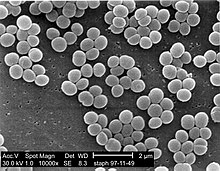
| Scanning electron micrograph of S. aureus colonies: Note the grape-like clustering common to Staphylococcus species. | |
| Scientific classification |
|
| Domain: | Bacteria |
| Phylum: | Bacillota |
| Class: | Bacilli |
| Order: | Bacillales |
| Family: | Staphylococcaceae |
| Genus: | Staphylococcus Rosenbach 1884 |
| Species | |
|
Staphylococcus is a genus of Gram-positive bacteria in the family Staphylococcaceae from the order Bacillales. Under the microscope, they appear spherical (cocci), and form in grape-like clusters. Staphylococcus species are facultative anaerobic organisms (capable of growth both aerobically and anaerobically).
The name was coined in 1880 by Scottish surgeon and bacteriologist Alexander Ogston (1844–1929), following the pattern established five years earlier with the naming of Streptococcus.[1] It combines the prefix «staphylo-» (from Ancient Greek: σταφυλή, romanized: staphylē, lit. ‘bunch of grapes’[2]), and suffixed by the Modern Latin: coccus, lit. ‘spherical bacterium’ (from Ancient Greek: κόκκος, romanized: kókkos, lit. ‘grain, seed, berry’[3]).
Staphylococcus was one of the leading infections in hospitals and many strains of this bacterium have become antibiotic resistant. Despite strong attempts to get rid of them, staph bacteria stay present in hospitals, where they can infect people who are most at risk of infection.[4]
Staphylococcus includes at least 43 species. Of these, nine have two subspecies, one has three subspecies, and one has four subspecies.[5] Many species cannot cause disease and reside normally on the skin and mucous membranes of humans and other animals. Staphylococcus species have been found to be nectar-inhabiting microbes.[6] They are also a small component of the soil microbiome.[7]
Taxonomy[edit]
The taxonomy is based on 16s rRNA sequences,[8] and most of the staphylococcal species fall into 11 clusters:
- S. aureus group – S. argenteus, S. aureus, S. schweitzeri, S. simiae
- S. auricularis group – S. auricularis
- S. carnosus group – S. carnosus, S. condimenti, S. debuckii, S. massiliensis, S. piscifermentans, S. simulans
- S. epidermidis group – S. capitis, S. caprae, S. epidermidis, S. saccharolyticus
- S. haemolyticus group – S. borealis, S. devriesei, S. haemolyticus, S. hominis
- S. hyicus-intermedius group – S. agnetis, S. chromogenes, S. cornubiensis, S. felis, S. delphini, S. hyicus, S. intermedius, S. lutrae, S. microti, S. muscae, S. pseudintermedius, S. rostri, S. schleiferi
- S. lugdunensis group – S. lugdunensis
- S. saprophyticus group – S. arlettae, S. caeli, S. cohnii, S. equorum, S. gallinarum, S. kloosii, S. leei, S. nepalensis, S. saprophyticus, S. succinus, S. xylosus
- S. sciuri group – S. fleurettii, S. lentus, S. sciuri, S. stepanovicii, S. vitulinus
- S. simulans group – S. simulans
- S. warneri group – S. pasteuri, S. warneri
A twelfth group – that of S. caseolyticus – has now been removed to a new genus, Macrococcus, the species of which are currently the closest known relatives of Staphylococcus.[9]
Two species were described in 2015 – Staphylococcus argenteus and Staphylococcus schweitzeri – both of which were previously considered variants of S. aureus.[10]
A new coagulase negative species – Staphylococcus edaphicus – has been isolated from Antarctica.[11] This species is probably a member of the S. saprophyticus group.
Groups[edit]
Based on an analysis of orthologous gene content three groups (A, B and C) have been proposed.[12]
Group A includes S. aureus, S. borealis, S. capitis, S. epidermidis, S. haemolyticus, S. hominis, S. lugdunensis, S. pettenkoferi, S. simiae and S. warneri.
Group B includes S. arlettae, S. cohnii, S. equorum, S. saprophyticus and S. xylosus.
Group C includes S. delphini, S. intermedius and S. pseudintermedius.
Notes[edit]
The S. saprophyticus and S. sciuri groups are generally novobiocin-resistant, as is S. hominis subsp. novobiosepticus.
Members of the S. sciuri group are oxidase-positive due to their possession of the enzyme cytochrome c oxidase. This group is the only clade within the staphylococci to possess this gene.
The S. sciuri group appears to be the closest relations to the genus Macrococcus.
S. pulvereri has been shown to be a junior synonym of S. vitulinus.[13]
Within these clades, the S. haemolyticus and S. simulans groups appear to be related, as do the S. aureus and S. epidermidis groups.[14]
S. lugdunensis appears to be related to the S. haemolyticus group.
S. petrasii may be related to S. haemolyticus, but this needs to be confirmed.
The taxonomic position of S. lyticans,S. petrasii, and S. pseudolugdunensis has yet to be clarified. The published descriptions of these species do not appear to have been validly published.
Biochemical identification[edit]
Assignment of a strain to the genus Staphylococcus requires it to be a Gram-positive coccus[15] that forms clusters, has an appropriate cell wall structure (including peptidoglycan type and teichoic acid presence) and G + C content of DNA in a range of 30–40 mol%.
Staphylococcus species can be differentiated from other aerobic and facultative anaerobic, Gram-positive cocci by several simple tests.[15] Staphylococcus species are facultative anaerobes (capable of growth both aerobically and anaerobically).[15] All species grow in the presence of bile salts.
All species of Staphylococcus aureus were once thought to be coagulase-positive, but this has since been disproven.[16][17][18]
Growth can also occur in a 6.5% NaCl solution.[15] On Baird-Parker medium, Staphylococcus species grow fermentatively, except for S. saprophyticus, which grows oxidatively. Staphylococcus species are resistant to bacitracin (0.04 U disc: resistance = < 10 mm zone of inhibition) and susceptible to furazolidone (100 μg disc: resistance = < 15 mm zone of inhibition). Further biochemical testing is needed to identify to the species level.
Coagulase production[edit]
One of the most important phenotypical features used in the classification of staphylococci is their ability to produce coagulase, an enzyme that causes blood clot formation.
Seven species are currently recognised as being coagulase-positive: S. aureus, S. delphini, S. hyicus, S. intermedius, S. lutrae, S. pseudintermedius, and S. schleiferi subsp. coagulans. These species belong to two separate groups – the S. aureus (S. aureus alone) group and the S. hyicus-intermedius group (the remaining five).
An eighth species has also been described – Staphylococcus leei – from patients with gastritis.[19]
S. aureus is coagulase-positive, meaning it produces coagulase. However, while the majority of S. aureus strains are coagulase-positive, some may be atypical in that they do not produce coagulase. S. aureus is catalase-positive (meaning that it can produce the enzyme catalase) and able to convert hydrogen peroxide (H2O2) to water and oxygen, which makes the catalase test useful to distinguish staphylococci from enterococci and streptococci.
S. pseudintermedius inhabits and sometimes infects the skin of domestic dogs and cats. This organism, too, can carry the genetic material that imparts multiple bacterial resistance. It is rarely implicated in infections in humans, as a zoonosis.
S. epidermidis, a coagulase-negative species, is a commensal of the skin, but can cause severe infections in immunosuppressed patients and those with central venous catheters.
S. saprophyticus, another coagulase-negative species that is part of the normal vaginal flora, is predominantly implicated in genitourinary tract infections in sexually active young women.
In recent years, several other Staphylococcus species have been implicated in human infections, notably S. lugdunensis, S. schleiferi, and S. caprae.
Common abbreviations for coagulase-negative staphylococci are CoNS, CNS, or CNST.[20] The American Society for Microbiology abbreviates coagulase-negative staphylococci as «CoNS».
Genomics and molecular biology[edit]
The first S. aureus genomes to be sequenced were those of N315 and Mu50, in 2001. Many more complete S. aureus genomes have been submitted to the public databases, making it one of the most extensively sequenced bacteria. The use of genomic data is now widespread and provides a valuable resource for researchers working with S. aureus. Whole genome technologies, such as sequencing projects and microarrays, have shown an enormous variety of S. aureus strains. Each contains different combinations of surface proteins and different toxins. Relating this information to pathogenic behaviour is one of the major areas of staphylococcal research. The development of molecular typing methods has enabled the tracking of different strains of S. aureus. This may lead to better control of outbreak strains. A greater understanding of how the staphylococci evolve, especially due to the acquisition of mobile genetic elements encoding resistance and virulence genes is helping to identify new outbreak strains and may even prevent their emergence.[21]
The widespread incidence of antibiotic resistance across various strains of S. aureus, or across different species of Staphylococcus has been attributed to horizontal gene transfer of genes encoding antibiotic/metal resistance and virulence. A recent study demonstrated the extent of horizontal gene transfer among Staphylococcus to be much greater than previously expected, and encompasses genes with functions beyond antibiotic resistance and virulence, and beyond genes residing within the mobile genetic elements.[22]
Various strains of Staphylococcus are available from biological research centres, such as the National Collection of Type Cultures.
Host range[edit]
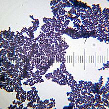
Unknown variety of Staphylococcus, Gram-stained – numbered ticks on the scale are 11 µm apart
Members of the genus Staphylococcus frequently colonize the skin and upper respiratory tracts of mammals and birds and also in marine sponge.[15] Marine sponge associated Staphylococcus species are highly salt tolerant.[15] Some species specificity has been observed in host range, such that the Staphylococcus species observed on some animals appear more rarely on more distantly related host species.[23]
Some of the observed host specificity includes:
- S. arlattae – chickens, goats, marine sponge[15]
- S. aureus – humans[24]
- S. auricularis – deer, dogs, humans
- S. borealis – humans, cattle
- S. capitis – humans
- S. caprae – goats, humans
- S. cohnii – chickens, humans
- S. delphini – dolphins
- S. devriesei – cattle
- S. epidermidis – humans, marine sponge[15]
- S. equorum – horses
- S. felis – cats
- S. fleurettii – goats
- S. gallinarum – chickens, goats, pheasants
- S. haemolyticus – humans, Cercocebus, Erythrocebus, Lemur, Macca, Microcebus, Pan
- S. hyicus – pigs
- S. leei – humans
- S. lentus – goats, rabbits, sheep
- S. lugdunensis – humans, goats
- S. lutrae – otters
- S. microti – voles (Microtus arvalis)
- S. nepalensis – goats
- S. pasteuri – humans, goats
- S. pettenkoferi – humans
- S. pseudintermedius – dogs
- S. rostri – pigs
- S. schleiferi – humans
- S. sciuri – humans, dogs, goats
- S. simiae – South American squirrel monkeys (Saimiri sciureus)
- S. simulans – humans
- S. warneri – humans, Cercopithecoidea, Pongidae
- S. xylosus – humans
Populations at risk for Staphylococcus aureus infection[edit]
It is said that anyone can develop a staph infection, although certain groups of people are at greater risk, including people with chronic conditions such as diabetes, cancer, vascular disease, eczema, lung disease, and people who inject drugs. In healthcare facilities, the risk of more serious staph infection is higher because many patients have weakened immune systems or have undergone procedures. In healthcare, the risk of more serious staph infection is higher for patients in intensive care units (ICUs), patients who have undergone certain types of surgeries and patients with medical devices inserted in their bodies.[25]
Clinical[edit]
Staphylococcus can cause a wide variety of diseases in humans and animals through either toxin production or penetration. Staphylococcal toxins are a common cause of food poisoning, for they can be produced by bacteria growing in improperly stored food items. The most common sialadenitis is caused by staphylococci, as bacterial infections.[26] Staphylococci break down leucine into isovaleric acid, the main odor of foot odor.[27]
See also[edit]
- Methicillin-resistant S. aureus (MRSA)
- Vancomycin-resistant S. aureus (VRSA)
References[edit]
- ^ «staphylococcus | Origin and meaning of staphylococcus by Online Etymology Dictionary». www.etymonline.com. Retrieved 2018-07-25.
- ^ stafulh/ in Liddell, Henry George; Scott, Robert (1940) A Greek–English Lexicon, revised and augmented throughout by Jones, Sir Henry Stuart, with the assistance of McKenzie, Roderick. Oxford: Clarendon Press. In the Perseus Digital Library, Tufts University.
- ^ ko)kkos in Liddell and Scott
- ^ «Staph infections». mayoclinic.org. Retrieved 2022-11-27.
- ^ Harris LG, Foster SJ, Richards RG (December 2002). «An introduction to Staphylococcus aureus, and techniques for identifying and quantifying S. aureus adhesins in relation to adhesion to biomaterials: review». European Cells & Materials. 4: 39–60. doi:10.22203/ecm.v004a04. PMID 14562246.
- ^ Jacquemyn H, Lenaerts M, Brys R, Willems K, Honnay O, Lievens B (2013). «Among-population variation in microbial community structure in the floral nectar of the bee-pollinated forest herb Pulmonaria officinalis L.» PLOS ONE. 8 (3): e56917. Bibcode:2013PLoSO…856917J. doi:10.1371/journal.pone.0056917. PMC 3594240. PMID 23536759.
- ^ Madigan M, Martinko J, eds. (2005). Brock Biomlogy of Microorganisms (11th ed.). Prentice Hall. ISBN 978-0-13-144329-7.[page needed]
- ^ Takahashi T, Satoh I, Kikuchi N (April 1999). «Phylogenetic relationships of 38 taxa of the genus Staphylococcus based on 16S rRNA gene sequence analysis». International Journal of Systematic Bacteriology. 49 (2): 725–8. doi:10.1099/00207713-49-2-725. PMID 10319495.
- ^ Kloos WE, Ballard DN, George CG, Webster JA, Hubner RJ, Ludwig W, Schleifer KH, Fiedler F, Schubert K (July 1998). «Delimiting the genus Staphylococcus through description of Macrococcus caseolyticus gen. nov., comb. nov. and Macrococcus equipercicus sp. nov., and Macrococcus bovicus sp. no. and Macrococcus carouselicus sp. nov». International Journal of Systematic Bacteriology. 48 (3): 859–77. doi:10.1099/00207713-48-3-859. PMID 9734040.
- ^ Tong SY, Schaumburg F, Ellington MJ, Corander J, Pichon B, Leendertz F, Bentley SD, Parkhill J, Holt DC, Peters G, Giffard PM (January 2015). «Novel staphylococcal species that form part of a Staphylococcus aureus-related complex: the non-pigmented Staphylococcus argenteus sp. nov. and the non-human primate-associated Staphylococcus schweitzeri sp. nov». International Journal of Systematic and Evolutionary Microbiology. 65 (Pt 1): 15–22. doi:10.1099/ijs.0.062752-0. PMC 4298100. PMID 25269845.
- ^ Pantůček R, Sedláček I, Indráková A, Vrbovská V, Mašlaňová I, Kovařovic V, Švec P, Králová S, Krištofová L, Kekláková J, Petráš P, Doškař J (October 2017). «mecCgene and genomic islands with suspected role in adaptation to extreme environment». Applied and Environmental Microbiology. 84 (2): e01746–17. doi:10.1128/AEM.01746-17. PMC 5752872. PMID 29079617.
- ^ Coates-Brown R, Moran JC, Pongchaikul P, Darby AC and MJ Horsburgh MJ (2018) «Comparative genomics of Staphylococcus reveals determinants of speciation and diversification of antimicrobial defense». Front Microbiol
- ^ Svec P, Vancanneyt M, Sedlácek I, Engelbeen K, Stetina V, Swings J, Petrás P (November 2004). «Reclassification of Staphylococcus pulvereri Zakrzewska-Czerwinska et al. 1995 as a later synonym of Staphylococcus vitulinus Webster et al. 1994″. International Journal of Systematic and Evolutionary Microbiology. 54 (Pt 6): 2213–5. doi:10.1099/ijs.0.63080-0. PMID 15545460.
- ^ Ghebremedhin B, Layer F, König W, König B (March 2008). «Genetic classification and distinguishing of Staphylococcus species based on different partial gap, 16S rRNA, hsp60, rpoB, sodA, and tuf gene sequences». Journal of Clinical Microbiology. 46 (3): 1019–25. doi:10.1128/JCM.02058-07. PMC 2268370. PMID 18174295.
- ^ a b c d e f g h Paul, Sulav Indra; Rahman, Md. Mahbubur; Salam, Mohammad Abdus; Khan, Md. Arifur Rahman; Islam, Md. Tofazzal (2021-12-15). «Identification of marine sponge-associated bacteria of the Saint Martin’s island of the Bay of Bengal emphasizing on the prevention of motile Aeromonas septicemia in Labeo rohita». Aquaculture. 545: 737156. doi:10.1016/j.aquaculture.2021.737156. ISSN 0044-8486.
- ^ Ryan KJ, Ray CG, eds. (2004). Sherris Medical Microbiology (4th ed.). McGraw Hill. ISBN 978-0-8385-8529-0.[page needed]
- ^ PreTest, Surgery, 12th ed., p. 88
- ^ Matthews KR, Roberson J, Gillespie BE, Luther DA, Oliver SP (1997). «Identification and Differentiation of Coagulase-Negative Staphylococcus aureus by Polymerase Chain Reaction». Journal of Food Protection. 60 (6): 686–8. doi:10.4315/0362-028X-60.6.686. PMID 31195568.
- ^ Jin M, Rosario W, Watler E, Calhoun DH (March 2004). «Development of a large-scale HPLC-based purification for the urease from Staphylococcus leei and determination of subunit structure». Protein Expression and Purification. 34 (1): 111–7. doi:10.1016/j.pep.2003.10.012. PMID 14766306.
- ^ Becker K, Heilmann C, Peters G (October 2014). «Coagulase-negative staphylococci». Clinical Microbiology Reviews. 27 (4): 870–926. doi:10.1128/CMR.00109-13. PMC 4187637. PMID 25278577.
- ^ Lindsay J, ed. (2008). Staphylococcus: Molecular Genetics. Caister Academic Press. ISBN 978-1-904455-29-5.[page needed]
- ^ Chan CX, Beiko RG, Ragan MA (August 2011). «Lateral transfer of genes and gene fragments in Staphylococcus extends beyond mobile elements». Journal of Bacteriology. 193 (15): 3964–77. doi:10.1128/JB.01524-10. PMC 3147504. PMID 21622749.
- ^ Kloos WE (1980). «Natural populations of the genus Staphylococcus«. Annual Review of Microbiology. 34: 559–92. doi:10.1146/annurev.mi.34.100180.003015. PMID 7002032.
- ^ «Symptoms and Treatments of Staph Infection (Cellulitis)». Just-Health.net. December 14, 2013.
- ^ «Staphylococcus aureus in Healthcare Settings | HAI | CDC». www.cdc.gov. 2020-12-10. Retrieved 2022-04-23.
- ^ «Sialoadenitis: inflammation of the salivary glands». The Medical Consumer’s Advocate. 2001-01-04. Retrieved 2011-01-04.
- ^ Stevens D, Cornmell R, Taylor D, Grimshaw SG, Riazanskaia S, Arnold DS, Fernstad SJ, Smith AM, Heaney LM, Reynolds JC, Thomas CL, Harker M. Spatial variations in the microbial community structure and diversity of the human foot is associated with the production of odorous volatiles. FEMS Microbiol Ecol. 2015 Jan;91(1):1-11. doi: 10.1093/femsec/fiu018. Epub 2014 Dec 8. PMID: 25764539.
External links[edit]
From Wikipedia, the free encyclopedia
«Staph» redirects here. Not to be confused with Staff.
| Staphylococcus | |
|---|---|
|
|
|
| Scanning electron micrograph of S. aureus colonies: Note the grape-like clustering common to Staphylococcus species. | |
| Scientific classification |
|
| Domain: | Bacteria |
| Phylum: | Bacillota |
| Class: | Bacilli |
| Order: | Bacillales |
| Family: | Staphylococcaceae |
| Genus: | Staphylococcus Rosenbach 1884 |
| Species | |
|
Staphylococcus is a genus of Gram-positive bacteria in the family Staphylococcaceae from the order Bacillales. Under the microscope, they appear spherical (cocci), and form in grape-like clusters. Staphylococcus species are facultative anaerobic organisms (capable of growth both aerobically and anaerobically).
The name was coined in 1880 by Scottish surgeon and bacteriologist Alexander Ogston (1844–1929), following the pattern established five years earlier with the naming of Streptococcus.[1] It combines the prefix «staphylo-» (from Ancient Greek: σταφυλή, romanized: staphylē, lit. ‘bunch of grapes’[2]), and suffixed by the Modern Latin: coccus, lit. ‘spherical bacterium’ (from Ancient Greek: κόκκος, romanized: kókkos, lit. ‘grain, seed, berry’[3]).
Staphylococcus was one of the leading infections in hospitals and many strains of this bacterium have become antibiotic resistant. Despite strong attempts to get rid of them, staph bacteria stay present in hospitals, where they can infect people who are most at risk of infection.[4]
Staphylococcus includes at least 43 species. Of these, nine have two subspecies, one has three subspecies, and one has four subspecies.[5] Many species cannot cause disease and reside normally on the skin and mucous membranes of humans and other animals. Staphylococcus species have been found to be nectar-inhabiting microbes.[6] They are also a small component of the soil microbiome.[7]
Taxonomy[edit]
The taxonomy is based on 16s rRNA sequences,[8] and most of the staphylococcal species fall into 11 clusters:
- S. aureus group – S. argenteus, S. aureus, S. schweitzeri, S. simiae
- S. auricularis group – S. auricularis
- S. carnosus group – S. carnosus, S. condimenti, S. debuckii, S. massiliensis, S. piscifermentans, S. simulans
- S. epidermidis group – S. capitis, S. caprae, S. epidermidis, S. saccharolyticus
- S. haemolyticus group – S. borealis, S. devriesei, S. haemolyticus, S. hominis
- S. hyicus-intermedius group – S. agnetis, S. chromogenes, S. cornubiensis, S. felis, S. delphini, S. hyicus, S. intermedius, S. lutrae, S. microti, S. muscae, S. pseudintermedius, S. rostri, S. schleiferi
- S. lugdunensis group – S. lugdunensis
- S. saprophyticus group – S. arlettae, S. caeli, S. cohnii, S. equorum, S. gallinarum, S. kloosii, S. leei, S. nepalensis, S. saprophyticus, S. succinus, S. xylosus
- S. sciuri group – S. fleurettii, S. lentus, S. sciuri, S. stepanovicii, S. vitulinus
- S. simulans group – S. simulans
- S. warneri group – S. pasteuri, S. warneri
A twelfth group – that of S. caseolyticus – has now been removed to a new genus, Macrococcus, the species of which are currently the closest known relatives of Staphylococcus.[9]
Two species were described in 2015 – Staphylococcus argenteus and Staphylococcus schweitzeri – both of which were previously considered variants of S. aureus.[10]
A new coagulase negative species – Staphylococcus edaphicus – has been isolated from Antarctica.[11] This species is probably a member of the S. saprophyticus group.
Groups[edit]
Based on an analysis of orthologous gene content three groups (A, B and C) have been proposed.[12]
Group A includes S. aureus, S. borealis, S. capitis, S. epidermidis, S. haemolyticus, S. hominis, S. lugdunensis, S. pettenkoferi, S. simiae and S. warneri.
Group B includes S. arlettae, S. cohnii, S. equorum, S. saprophyticus and S. xylosus.
Group C includes S. delphini, S. intermedius and S. pseudintermedius.
Notes[edit]
The S. saprophyticus and S. sciuri groups are generally novobiocin-resistant, as is S. hominis subsp. novobiosepticus.
Members of the S. sciuri group are oxidase-positive due to their possession of the enzyme cytochrome c oxidase. This group is the only clade within the staphylococci to possess this gene.
The S. sciuri group appears to be the closest relations to the genus Macrococcus.
S. pulvereri has been shown to be a junior synonym of S. vitulinus.[13]
Within these clades, the S. haemolyticus and S. simulans groups appear to be related, as do the S. aureus and S. epidermidis groups.[14]
S. lugdunensis appears to be related to the S. haemolyticus group.
S. petrasii may be related to S. haemolyticus, but this needs to be confirmed.
The taxonomic position of S. lyticans,S. petrasii, and S. pseudolugdunensis has yet to be clarified. The published descriptions of these species do not appear to have been validly published.
Biochemical identification[edit]
Assignment of a strain to the genus Staphylococcus requires it to be a Gram-positive coccus[15] that forms clusters, has an appropriate cell wall structure (including peptidoglycan type and teichoic acid presence) and G + C content of DNA in a range of 30–40 mol%.
Staphylococcus species can be differentiated from other aerobic and facultative anaerobic, Gram-positive cocci by several simple tests.[15] Staphylococcus species are facultative anaerobes (capable of growth both aerobically and anaerobically).[15] All species grow in the presence of bile salts.
All species of Staphylococcus aureus were once thought to be coagulase-positive, but this has since been disproven.[16][17][18]
Growth can also occur in a 6.5% NaCl solution.[15] On Baird-Parker medium, Staphylococcus species grow fermentatively, except for S. saprophyticus, which grows oxidatively. Staphylococcus species are resistant to bacitracin (0.04 U disc: resistance = < 10 mm zone of inhibition) and susceptible to furazolidone (100 μg disc: resistance = < 15 mm zone of inhibition). Further biochemical testing is needed to identify to the species level.
Coagulase production[edit]
One of the most important phenotypical features used in the classification of staphylococci is their ability to produce coagulase, an enzyme that causes blood clot formation.
Seven species are currently recognised as being coagulase-positive: S. aureus, S. delphini, S. hyicus, S. intermedius, S. lutrae, S. pseudintermedius, and S. schleiferi subsp. coagulans. These species belong to two separate groups – the S. aureus (S. aureus alone) group and the S. hyicus-intermedius group (the remaining five).
An eighth species has also been described – Staphylococcus leei – from patients with gastritis.[19]
S. aureus is coagulase-positive, meaning it produces coagulase. However, while the majority of S. aureus strains are coagulase-positive, some may be atypical in that they do not produce coagulase. S. aureus is catalase-positive (meaning that it can produce the enzyme catalase) and able to convert hydrogen peroxide (H2O2) to water and oxygen, which makes the catalase test useful to distinguish staphylococci from enterococci and streptococci.
S. pseudintermedius inhabits and sometimes infects the skin of domestic dogs and cats. This organism, too, can carry the genetic material that imparts multiple bacterial resistance. It is rarely implicated in infections in humans, as a zoonosis.
S. epidermidis, a coagulase-negative species, is a commensal of the skin, but can cause severe infections in immunosuppressed patients and those with central venous catheters.
S. saprophyticus, another coagulase-negative species that is part of the normal vaginal flora, is predominantly implicated in genitourinary tract infections in sexually active young women.
In recent years, several other Staphylococcus species have been implicated in human infections, notably S. lugdunensis, S. schleiferi, and S. caprae.
Common abbreviations for coagulase-negative staphylococci are CoNS, CNS, or CNST.[20] The American Society for Microbiology abbreviates coagulase-negative staphylococci as «CoNS».
Genomics and molecular biology[edit]
The first S. aureus genomes to be sequenced were those of N315 and Mu50, in 2001. Many more complete S. aureus genomes have been submitted to the public databases, making it one of the most extensively sequenced bacteria. The use of genomic data is now widespread and provides a valuable resource for researchers working with S. aureus. Whole genome technologies, such as sequencing projects and microarrays, have shown an enormous variety of S. aureus strains. Each contains different combinations of surface proteins and different toxins. Relating this information to pathogenic behaviour is one of the major areas of staphylococcal research. The development of molecular typing methods has enabled the tracking of different strains of S. aureus. This may lead to better control of outbreak strains. A greater understanding of how the staphylococci evolve, especially due to the acquisition of mobile genetic elements encoding resistance and virulence genes is helping to identify new outbreak strains and may even prevent their emergence.[21]
The widespread incidence of antibiotic resistance across various strains of S. aureus, or across different species of Staphylococcus has been attributed to horizontal gene transfer of genes encoding antibiotic/metal resistance and virulence. A recent study demonstrated the extent of horizontal gene transfer among Staphylococcus to be much greater than previously expected, and encompasses genes with functions beyond antibiotic resistance and virulence, and beyond genes residing within the mobile genetic elements.[22]
Various strains of Staphylococcus are available from biological research centres, such as the National Collection of Type Cultures.
Host range[edit]
Unknown variety of Staphylococcus, Gram-stained – numbered ticks on the scale are 11 µm apart
Members of the genus Staphylococcus frequently colonize the skin and upper respiratory tracts of mammals and birds and also in marine sponge.[15] Marine sponge associated Staphylococcus species are highly salt tolerant.[15] Some species specificity has been observed in host range, such that the Staphylococcus species observed on some animals appear more rarely on more distantly related host species.[23]
Some of the observed host specificity includes:
- S. arlattae – chickens, goats, marine sponge[15]
- S. aureus – humans[24]
- S. auricularis – deer, dogs, humans
- S. borealis – humans, cattle
- S. capitis – humans
- S. caprae – goats, humans
- S. cohnii – chickens, humans
- S. delphini – dolphins
- S. devriesei – cattle
- S. epidermidis – humans, marine sponge[15]
- S. equorum – horses
- S. felis – cats
- S. fleurettii – goats
- S. gallinarum – chickens, goats, pheasants
- S. haemolyticus – humans, Cercocebus, Erythrocebus, Lemur, Macca, Microcebus, Pan
- S. hyicus – pigs
- S. leei – humans
- S. lentus – goats, rabbits, sheep
- S. lugdunensis – humans, goats
- S. lutrae – otters
- S. microti – voles (Microtus arvalis)
- S. nepalensis – goats
- S. pasteuri – humans, goats
- S. pettenkoferi – humans
- S. pseudintermedius – dogs
- S. rostri – pigs
- S. schleiferi – humans
- S. sciuri – humans, dogs, goats
- S. simiae – South American squirrel monkeys (Saimiri sciureus)
- S. simulans – humans
- S. warneri – humans, Cercopithecoidea, Pongidae
- S. xylosus – humans
Populations at risk for Staphylococcus aureus infection[edit]
It is said that anyone can develop a staph infection, although certain groups of people are at greater risk, including people with chronic conditions such as diabetes, cancer, vascular disease, eczema, lung disease, and people who inject drugs. In healthcare facilities, the risk of more serious staph infection is higher because many patients have weakened immune systems or have undergone procedures. In healthcare, the risk of more serious staph infection is higher for patients in intensive care units (ICUs), patients who have undergone certain types of surgeries and patients with medical devices inserted in their bodies.[25]
Clinical[edit]
Staphylococcus can cause a wide variety of diseases in humans and animals through either toxin production or penetration. Staphylococcal toxins are a common cause of food poisoning, for they can be produced by bacteria growing in improperly stored food items. The most common sialadenitis is caused by staphylococci, as bacterial infections.[26] Staphylococci break down leucine into isovaleric acid, the main odor of foot odor.[27]
See also[edit]
- Methicillin-resistant S. aureus (MRSA)
- Vancomycin-resistant S. aureus (VRSA)
References[edit]
- ^ «staphylococcus | Origin and meaning of staphylococcus by Online Etymology Dictionary». www.etymonline.com. Retrieved 2018-07-25.
- ^ stafulh/ in Liddell, Henry George; Scott, Robert (1940) A Greek–English Lexicon, revised and augmented throughout by Jones, Sir Henry Stuart, with the assistance of McKenzie, Roderick. Oxford: Clarendon Press. In the Perseus Digital Library, Tufts University.
- ^ ko)kkos in Liddell and Scott
- ^ «Staph infections». mayoclinic.org. Retrieved 2022-11-27.
- ^ Harris LG, Foster SJ, Richards RG (December 2002). «An introduction to Staphylococcus aureus, and techniques for identifying and quantifying S. aureus adhesins in relation to adhesion to biomaterials: review». European Cells & Materials. 4: 39–60. doi:10.22203/ecm.v004a04. PMID 14562246.
- ^ Jacquemyn H, Lenaerts M, Brys R, Willems K, Honnay O, Lievens B (2013). «Among-population variation in microbial community structure in the floral nectar of the bee-pollinated forest herb Pulmonaria officinalis L.» PLOS ONE. 8 (3): e56917. Bibcode:2013PLoSO…856917J. doi:10.1371/journal.pone.0056917. PMC 3594240. PMID 23536759.
- ^ Madigan M, Martinko J, eds. (2005). Brock Biomlogy of Microorganisms (11th ed.). Prentice Hall. ISBN 978-0-13-144329-7.[page needed]
- ^ Takahashi T, Satoh I, Kikuchi N (April 1999). «Phylogenetic relationships of 38 taxa of the genus Staphylococcus based on 16S rRNA gene sequence analysis». International Journal of Systematic Bacteriology. 49 (2): 725–8. doi:10.1099/00207713-49-2-725. PMID 10319495.
- ^ Kloos WE, Ballard DN, George CG, Webster JA, Hubner RJ, Ludwig W, Schleifer KH, Fiedler F, Schubert K (July 1998). «Delimiting the genus Staphylococcus through description of Macrococcus caseolyticus gen. nov., comb. nov. and Macrococcus equipercicus sp. nov., and Macrococcus bovicus sp. no. and Macrococcus carouselicus sp. nov». International Journal of Systematic Bacteriology. 48 (3): 859–77. doi:10.1099/00207713-48-3-859. PMID 9734040.
- ^ Tong SY, Schaumburg F, Ellington MJ, Corander J, Pichon B, Leendertz F, Bentley SD, Parkhill J, Holt DC, Peters G, Giffard PM (January 2015). «Novel staphylococcal species that form part of a Staphylococcus aureus-related complex: the non-pigmented Staphylococcus argenteus sp. nov. and the non-human primate-associated Staphylococcus schweitzeri sp. nov». International Journal of Systematic and Evolutionary Microbiology. 65 (Pt 1): 15–22. doi:10.1099/ijs.0.062752-0. PMC 4298100. PMID 25269845.
- ^ Pantůček R, Sedláček I, Indráková A, Vrbovská V, Mašlaňová I, Kovařovic V, Švec P, Králová S, Krištofová L, Kekláková J, Petráš P, Doškař J (October 2017). «mecCgene and genomic islands with suspected role in adaptation to extreme environment». Applied and Environmental Microbiology. 84 (2): e01746–17. doi:10.1128/AEM.01746-17. PMC 5752872. PMID 29079617.
- ^ Coates-Brown R, Moran JC, Pongchaikul P, Darby AC and MJ Horsburgh MJ (2018) «Comparative genomics of Staphylococcus reveals determinants of speciation and diversification of antimicrobial defense». Front Microbiol
- ^ Svec P, Vancanneyt M, Sedlácek I, Engelbeen K, Stetina V, Swings J, Petrás P (November 2004). «Reclassification of Staphylococcus pulvereri Zakrzewska-Czerwinska et al. 1995 as a later synonym of Staphylococcus vitulinus Webster et al. 1994″. International Journal of Systematic and Evolutionary Microbiology. 54 (Pt 6): 2213–5. doi:10.1099/ijs.0.63080-0. PMID 15545460.
- ^ Ghebremedhin B, Layer F, König W, König B (March 2008). «Genetic classification and distinguishing of Staphylococcus species based on different partial gap, 16S rRNA, hsp60, rpoB, sodA, and tuf gene sequences». Journal of Clinical Microbiology. 46 (3): 1019–25. doi:10.1128/JCM.02058-07. PMC 2268370. PMID 18174295.
- ^ a b c d e f g h Paul, Sulav Indra; Rahman, Md. Mahbubur; Salam, Mohammad Abdus; Khan, Md. Arifur Rahman; Islam, Md. Tofazzal (2021-12-15). «Identification of marine sponge-associated bacteria of the Saint Martin’s island of the Bay of Bengal emphasizing on the prevention of motile Aeromonas septicemia in Labeo rohita». Aquaculture. 545: 737156. doi:10.1016/j.aquaculture.2021.737156. ISSN 0044-8486.
- ^ Ryan KJ, Ray CG, eds. (2004). Sherris Medical Microbiology (4th ed.). McGraw Hill. ISBN 978-0-8385-8529-0.[page needed]
- ^ PreTest, Surgery, 12th ed., p. 88
- ^ Matthews KR, Roberson J, Gillespie BE, Luther DA, Oliver SP (1997). «Identification and Differentiation of Coagulase-Negative Staphylococcus aureus by Polymerase Chain Reaction». Journal of Food Protection. 60 (6): 686–8. doi:10.4315/0362-028X-60.6.686. PMID 31195568.
- ^ Jin M, Rosario W, Watler E, Calhoun DH (March 2004). «Development of a large-scale HPLC-based purification for the urease from Staphylococcus leei and determination of subunit structure». Protein Expression and Purification. 34 (1): 111–7. doi:10.1016/j.pep.2003.10.012. PMID 14766306.
- ^ Becker K, Heilmann C, Peters G (October 2014). «Coagulase-negative staphylococci». Clinical Microbiology Reviews. 27 (4): 870–926. doi:10.1128/CMR.00109-13. PMC 4187637. PMID 25278577.
- ^ Lindsay J, ed. (2008). Staphylococcus: Molecular Genetics. Caister Academic Press. ISBN 978-1-904455-29-5.[page needed]
- ^ Chan CX, Beiko RG, Ragan MA (August 2011). «Lateral transfer of genes and gene fragments in Staphylococcus extends beyond mobile elements». Journal of Bacteriology. 193 (15): 3964–77. doi:10.1128/JB.01524-10. PMC 3147504. PMID 21622749.
- ^ Kloos WE (1980). «Natural populations of the genus Staphylococcus«. Annual Review of Microbiology. 34: 559–92. doi:10.1146/annurev.mi.34.100180.003015. PMID 7002032.
- ^ «Symptoms and Treatments of Staph Infection (Cellulitis)». Just-Health.net. December 14, 2013.
- ^ «Staphylococcus aureus in Healthcare Settings | HAI | CDC». www.cdc.gov. 2020-12-10. Retrieved 2022-04-23.
- ^ «Sialoadenitis: inflammation of the salivary glands». The Medical Consumer’s Advocate. 2001-01-04. Retrieved 2011-01-04.
- ^ Stevens D, Cornmell R, Taylor D, Grimshaw SG, Riazanskaia S, Arnold DS, Fernstad SJ, Smith AM, Heaney LM, Reynolds JC, Thomas CL, Harker M. Spatial variations in the microbial community structure and diversity of the human foot is associated with the production of odorous volatiles. FEMS Microbiol Ecol. 2015 Jan;91(1):1-11. doi: 10.1093/femsec/fiu018. Epub 2014 Dec 8. PMID: 25764539.
External links[edit]
Стафилококк
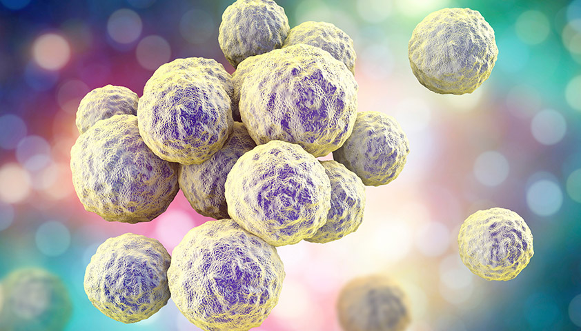
Стафилококки — это бактерии круглой формы, способные жить и размножаться в бескислородной среде и создавать колонии, похожие под микроскопом на гроздья винограда. Золотистый, эпидермальный и сапрофитный стафилококк — возбудители инфекционных заболеваний.
СОДЕРЖАНИЕ
Виды стафилококковых инфекций
Причины возникновения стафилококковых инфекций
Симптомы стафилококковой инфекции у взрослых
Стафилококковая инфекция при беременности
Стафилококковая инфекция у детей
Диагностика стафилококковой инфекции
Лечение стафилококковой инфекции
Профилактика стафилококковой инфекции
Что такое стафилококк
Стафилококки — бактерии, которым не нужен кислород для жизни и размножения.
Всего известно около 40 видов стафилококков. Большинство из них безвредны для человека. Три вида — золотистый, эпидермальный и сапрофитный стафилококк — могут стать причиной инфекционных заболеваний.
- Сапрофитный стафилококк (S. saprophyticus) чаще всего обитает в области гениталий и мочеиспускательного канала (уретры). Может спровоцировать цистит, уретрит.
- Эпидермальный (S. epidermidis) селится на любых участках кожи и слизистых оболочках. Наиболее опасен для пациентов после операций, связанных с внутренним протезированием: стафилококк может попасть внутрь вместе с протезом (шунтом, клапаном) и спровоцировать эндокардит — воспаление внутренней оболочки сердца, суставные инфекции, воспаление мочевыводящих путей.
- Золотистый стафилококк (S. aureus) — наиболее агрессивный представитель своего рода. Проникнув во внутреннюю среду организма, он может вызвать воспалительный процесс в органах и тканях. Устойчив к антибиотикам и антисептикам. Хорошо выживает в неблагоприятной среде: не теряет активности при высушивании, 12 часов может находиться под прямыми солнечными лучами, в течение 10 минут выдерживает температуру 150 °С, не погибает в этиловом спирте и перекиси водорода.
Название «стафилококк» происходит от древнегреческих слов σταφυλή («гроздь винограда») и κόκκος («зерно, ягода»). Колония этих бактерий под микроскопом действительно похожа на виноградную кисть.
Как можно заразиться стафилококком
Есть несколько путей передачи стафилококка:
- Контактно-бытовой. Стафилококк может попасть в организм через заражённые предметы: дверные ручки, средства личной гигиены.
- Воздушно-капельный. Бактерия выделяется во внешнюю среду, когда заражённый человек кашляет, чихает и даже разговаривает.
- Пищевой. Стафилококк передаётся через сырую воду, загрязнённые продукты, непромытую посуду.
- Вертикальный. Ребёнок может заразиться стафилококком от матери во время родов.
Стафилококковое пищевое отравление можно получить, если съесть просроченный продукт, особенно йогурт, кефир, простоквашу.
Виды стафилококковых инфекций
Стафилококковая инфекция — группа гнойно-воспалительных заболеваний, вызванных патогенными штаммами бактерий.
Инфекции, связанные с прямым проникновением стафилококка в ткани и органы:
- локализованные кожные инфекции: ячмень, абсцессы, фурункулы, карбункулы, фолликулиты;
- диффузные кожные инфекции: вульгарное импетиго — гнойничковое заболевание кожи; целлюлит (гнойное воспаление подкожной жировой клетчатки), в том числе орбитальный целлюлит — воспаление тканей глаза за орбитальной перегородкой;
- инфекции внутренних органов и тканей: септический артрит — воспаление сустава; острый и хронический остеомиелит — воспаление костного мозга; сепсис; пневмония; менингит; эндокардит — воспаление внутренней оболочки сердца;
- другие инфекции: астма, ринит, синусит.
Токсикозы — состояния, вызванные стафилококковым токсином:
- гастроэнтерит (пищевое отравление) — воспаление желудка и тонкого кишечника;
- синдром стафилококкового токсического шока (СТШ);
- стафилококковый синдром ошпаренной кожи (ССОК) — заболевание, при котором на коже образуются пузыри с отслойкой эпидермиса.
Причины возникновения стафилококковых инфекций
У большинства людей сапрофитный, эпидермальный и золотистый стафилококк в норме входят в состав микрофлоры и никак себя не проявляют. Иммунная система замедляет рост бактерий и удерживает их численность в безопасных пределах.
Но из-за снижения иммунитета на фоне болезней, стресса, неправильного питания может развиться стафилококковая инфекция.
Группы риска по развитию стафилококковой инфекции:
- пациенты с иммунодефицитом, в том числе вызванным ВИЧ — вирусом иммунодефицита человека;
- пациенты с сахарным диабетом, эндокринными заболеваниями, хроническими бронхолёгочными болезнями (например, с бронхиальной астмой);
- пациенты с имплантированными протезами;
- пациенты, которые проходят лечение кортикоидами (стероидными гормонами), иммунодепрессантами (препаратами, искусственно угнетающими иммунитет);
- пациенты на диализе;
- новорождённые, дети в первые годы жизни (в среднем до 5 лет);
- беременные;
- люди старше 60 лет;
- курильщики, люди, злоупотребляющие алкоголем.
Симптомы стафилококковой инфекции у взрослых
Симптомы инфекции зависят от локализации колонии бактерий.
Стафилококковая инфекция кожи — наиболее распространённая форма заболевания. Основные проявления: узелки, пустулы, ссыхающиеся в корки. Более глубокие поражения: фурункул, карбункул или абсцесс кожи в виде болезненного узла, с последующим формированием полости, заполненной гноем.
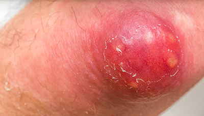
Карбункул — конгломерат фурункулов, объединённых общим инфильтратом
Стафилококковое пищевое отравление связано с употреблением заражённых продуктов. Многие продукты служат питательной средой для бактерий, сохраняя при этом нормальный запах и вкус.
Как правило, через 2–8 часов после еды появляется тяжёлая тошнота, рвота, боль в животе, диарея.
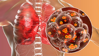
Стафилококковая пневмония — не самое распространённое проявление стафилококковой инфекции, но она может развиться у пациентов, которые болеют или переболели гриппом, принимают кортикостероиды, иммунодепрессанты, а также у людей с хроническими бронхолёгочными заболеваниями.
Симптомы стафилококковой пневмонии:
- общее недомогание,
- выраженная интоксикация,
- высокая температура,
- озноб,
- одышка,
- болезненный кашель.
Стафилококковый эндокардит — воспаление внутренней оболочки сердца
Стафилококковый эндокардит — воспаление внутренней оболочки сердца.
Симптомы бактериального эндокардита:
- слабость,
- одышка,
- лихорадка,
- плохой аппетит,
- потеря веса,
- озноб,
- бледность кожи и слизистых оболочек.
Если стафилококковый эндокардит не начать лечить вовремя, могут развиться осложнения: септический шок, эмболия (перекрытие просвета) сосудов сердца и головного мозга, сердечная недостаточность.
Стафилококковый артрит — воспалительное заболевание, которое часто развивается после операции на суставах и установки суставного протеза.
Местные проявления стафилококкового артрита: отёк и покраснение кожи в области поражённого сустава, резкая боль, ограничение движения.
Общие проявления стафилококкового артрита: повышенная температура, озноб, выраженная интоксикация. У детей встречается также тошнота и рвота.
Инфекционный артрит, вызванный золотистым стафилококком, может привести к разрушению хряща сустава за несколько дней.
Стафилококковая инфекция при беременности
Период беременности может сопровождаться снижением иммунитета и обострением хронических заболеваний — а это создаёт благоприятные условия для роста колоний стафилококков.
Сапрофитный стафилококк не считается особенно опасным для беременных женщин. Как правило, он локализуется в органах мочеполовой системы и почках и провоцирует развитие цистита, уретрита, пиелонефрита.
Благодаря выраженной симптоматике, сапрофитного стафилококка достаточно легко обнаружить в бактериальном посеве и вовремя принять меры, чтобы не допустить дальнейшего развития инфекции и осложнений.
Эпидермальный стафилококк достаточно быстро распространяется по кровотоку и может проникнуть через плацентарный барьер от матери к плоду, приводя к нарушениям его роста и развития.
Золотистый стафилококк — самый опасный. Если он проникает во внутреннюю среду организма, может развиться сепсис, что приведёт к поражению нервной системы и внутренних органов эмбриона. Также заражение ребёнка стафилококком во время родов повышает риск развития у него инфекции кожи и мягких тканей.
Стафилококковая инфекция у детей
У новорождённых стафилококк вызывает ряд специфических заболеваний:
- Эксфолиативный дерматит Риттера — тяжёлое инфекционное поражение кожи новорождённых, при котором она краснеет, образуются пузыри.
- Синдром ошпаренной кожи — острое поражение кожного покрова, вызванное одним из токсинов стафилококка. Характеризуется покраснением, отслойкой эпидермиса с образованием пузырей, напоминает ожог II степени.
- Эпидемическая пузырчатка новорождённых — острое инфекционное заболевание, при котором на коже образуются пузыри, заполненные серозным содержимым, и лопаются даже при небольшом касании. Инфицированная жидкость попадает на кожу и провоцирует появление новых пузырей.
- Острый мастит — гнойно-воспалительное заболевание грудной железы. Обычно проявляется на 7–10-й день жизни ребёнка. Сопровождается уплотнением железы (обычно с одной стороны), повышением температуры до 39 С°.
В дошкольном и школьном возрасте инфицирование стафилококком может привести к развитию синусита, ринита, фарингита, бронхита и пневмонии.
Также достаточно распространены поражения кожи, причём у детей их может вызвать не только золотистый стафилококк, но и эпидермальный, сапрофитный — как правило, при иммунодефицитных состояниях.
Диагностика стафилококковой инфекции
К какому врачу обращаться
Лечением стафилококковой инфекции занимаются врачи-инфекционисты. Если нет точно поставленного диагноза, можно обратиться к терапевту или педиатру для обследования.
Какие анализы нужно сдать
Анализ на стафилококк берут из той зоны, в которой предположительно развивается инфекция.
Современные методы позволяют исследовать самые разные биоматериалы и биологические жидкости: грудное молоко, кал, мочу, мокроту, соскоб из зева, носа или с конъюнктивы, отделяемое из уха и раневое отделяемое.
141.0. Взятие (2 вида, +450 ₽)  95 Колич. 2 дня
Взятие (2 вида, +450 ₽) Колич. 2 дня
 95 бонусов на счёт
Соскоб кожи проверяют при дерматологических проблемах. Мазок из горла и носа — если есть симптомы поражения верхних дыхательных путей. Кал исследуют при расстройствах пищеварения, мочу — при симптомах цистита. Кровь на анализ берут, если есть подозрение на генерализацию инфекции: чтобы определить присутствие бактерий в крови.
141.0.01.16.01.0. Соскоб (+450 ₽) 125 3 дня
Соскоб (+450 ₽) 3 дня
125 бонусов на счёт
141.0.01.10.01.0. Соскоб (+450 ₽) 130 3 дня
Соскоб (+450 ₽) 3 дня
130 бонусов на счёт
120.2. Соскоб (+450 ₽) 110 4 дня
Соскоб (+450 ₽) 4 дня
110 бонусов на счёт
141.0.05.19.01.0. 150 3 дня
3 дня
150 бонусов на счёт
124.0. 198 4 дня
4 дня
198 бонусов на счёт
Концентрация бактерий, которую можно считать нормальной, зависит от возраста пациента, его медицинского анамнеза и состояния организма. Так, количество стафилококков, которое не приносит вреда взрослому человеку с нормальным иммунитетом, может представлять серьёзную угрозу для новорождённого.
Лечение стафилококковой инфекции
Для лечения стафилококковой инфекции применяют антибиотики нового поколения — полусинтетические пенициллины, аминогликозиды и некоторые другие. Также используют бактериофаги — специфические вирусы, которые прицельно уничтожают именно стафилококки. При инфекционных заболеваниях кожи назначают средства с антимикробными компонентами, антисептики.
Профилактика стафилококковой инфекции
Меры профилактики стафилококковой инфекции:
- избегать переохлаждения;
- следить за состоянием иммунной системы;
- поддерживать баланс витаминов в организме;
- соблюдать правила личной гигиены;
- избегать контакта с заражёнными людьми;
- термически обрабатывать сырое мясо.
Такие несложные меры профилактики помогут избежать стафилококковой инфекции.
Частые вопросы
Чаще всего стафилококковая инфекция поражает кожу. Стафилококк может вызывать ячмень, абсцессы, фурункулы, карбункулы и фолликулиты, а также вульгарное импетиго и целлюлит — гнойное воспаление подкожной жировой клетчатки.
Если бактерия поражает внутренние органы и ткани, может развиться септический артрит (воспаление сустава), остеомиелит (воспаление костного мозга), эндокардит (воспаление внутренней оболочки сердца), сепсис (заражение крови), пневмония, менингит. При поражении органов дыхания возникают астма, ринит и синусит.
Выделяемые бактерией токсины могут вызвать гастроэнтерит (воспаление желудка и тонкого кишечника) и синдром стафилококкового токсического шока.
Если симптомов нет, то лечение не требуется, так как стафилококк — часть нормальной микрофлоры человека. Если развивается воспалительная патология, например синусит, нужно обратиться к отоларингологу — врач подберёт оптимальное лечение.
Основной способ выявления стафилококковой инфекции — лабораторная диагностика. Современные методы позволяют исследовать самые разные биоматериалы и биологические жидкости: грудное молоко, кал, мочу, мокроту, раневое отделяемое и т.д.
Анализ на стафилококк берут из той зоны, в которой предположительно развивается инфекция. Соскоб кожи проверяют при дерматологических проблемах. Мазок из горла и носа — если есть симптомы поражения верхних дыхательных путей. Кал исследуют при расстройствах пищеварения, мочу — при симптомах цистита. Кровь на анализ берут, если есть подозрение на генерализацию инфекции — чтобы определить присутствие бактерий в крови.
Есть несколько способов передачи стафилококка: контактно-бытовой, воздушно-капельный, пищевой, вертикальный — от матери к ребёнку во время родов.
Продолжительность лечения — от 7 дней до нескольких месяцев, в зависимости от тяжести и формы заболевания.
Информацию проверил
врач-эксперт

Информацию проверил врач-эксперт
Роман Иванов
Врач-дерматовенеролог
Оцените статью:
Полезная статья? Поделитесь в социальных сетях:
ВАЖНО
Информация из данного раздела не может служить достаточным основанием для постановки диагноза или назначения лечения. Решение об этом должен принимать врач на основании всех имеющихся у него данных.
Вам может быть интересно
Вам телеграм.
Telegram-канал,
которому, на наш взгляд,
можно доверять