Запрос «ОДА» перенаправляется сюда; см. также другие значения.
Опорно-двигательная система (синонимы: опорно-двигательный аппарат, костно-мышечная система, локомоторная система, скелетно-мышечная система) — комплекс структур, образующих каркас, придающий форму организму, дающий ему опору, обеспечивающий защиту внутренних органов и возможность передвижения в пространстве[1].
Опорно-двигательная система человека — функциональная совокупность костей скелета, их соединений (суставов и синартрозов), и соматической мускулатуры со вспомогательными приспособлениями, осуществляющих посредством нервной регуляции локомоции, поддержание позы, мимики и других двигательных действиях, наряду с другими системами органов образует человеческое тело.
В англоязычной литературе применяют близкие по значению термины: скелетно-мышечная система и локомоторная система.
Содержание
- 1 Науки, изучающие опорно-двигательную систему человека
- 2 Функциональная анатомия
- 3 Функции двигательного аппарата
- 4 См. также
- 5 Примечания
- 6 См. также
Науки, изучающие опорно-двигательную систему человека
- медицина
- анатомия
- физиология (физиология движений)
- ортопедия
- травматология
- спортивная медицина
- протезирование
- клиническая биомеханика
- биомеханика
- физическая культура
- теория физической культуры
- биомеханика физических упражнений
Функциональная анатомия
Двигательный аппарат человека — это самодвижущийся механизм, состоящий из 400 мышц, 206 костей, нескольких сотен сухожилий. Опорно-двигательная система человека — функциональная совокупность костей скелета, их соединений (суставов и синартрозов), и соматической мускулатуры и других двигательных действиях, наряду с другими системами органов образует человеческое тело.
Функции двигательного аппарата
- опорная — фиксация мышц и внутренних органов;
- защитная — защита жизненно важных органов (головной мозг и спинной мозг, сердце и др.);
- двигательная — обеспечение простых движений, двигательных действий (осанка, локомоции, манипуляции) и двигательной деятельности;
- рессорная — смягчение толчков и сотрясений;
- биологическая — участие в обеспечении жизненно важных процессов, такие как минеральный обмен, кровообращение, кроветворение и другие.
Двигательная функция возможна только при условии взаимодействия костей и мышц скелета, потому что мышцы приводят в движение костные рычаги. Большинство костей скелета соединено подвижно с помощью суставов. Одним концом мышца прикрепляется к одной кости, образуя сустав, другим концом — к другой кости. При сокращении мышца приводит кости в движение. Благодаря мышцам противоположного действия кости могут не только совершать те или иные движения, но и фиксироваться относительно друг друга.
Кости и мышцы принимают участие в обмене веществ, в частности в обмене кальция и фосфора.
См. также
- Заболевания опорно-двигательного аппарата человека
- Позвоночник человека
- Осанка
- Биомеханика
- Моторика
Примечания
- ↑ Статья «Опорно-двигательная система» в словаре по естественным наукам «Глоссарий.ру»
См. также
| |
|---|
| Сердечно-сосудистая система (сердце, сосуды) • Лимфатическая система • Пищеварительная система • Эндокринная система • Иммунная система • Сенсорная система (соматосенсорная система, зрительная система, обонятельная сенсорная система, слуховая сенсорная система, вкусовая сенсорная система) • Покровная система • Нервная система (центральная, периферическая) • Опорно-двигательная система (костная система, мышечная система) • мочеполовая система (репродуктивная система, мочевыделительная система) • Дыхательная система |
| |
|
|---|---|
| Экзоскелет |
Неорганический: Раковина (многих простейших, моллюсков) Органический: Кутикула (хитиновый панцирь членистоногих) |
| Эндоскелет |
Осевой (позвоночный столб (позвоночник) • череп • грудная клетка) Добавочный (пояс верхних конечностей • верхние конечности • пояс нижних конечностей • нижние конечности) |
| Гидроскелет | Образование восьмилучевых полипов |
| См. также | Скелетная революция • Скелет человека • Перечень костей скелета человека • Опорно-двигательная система |
From Wikipedia, the free encyclopedia
| Musculoskeletal system | |
|---|---|

Features of the human activity system from the 1911 Encyclopædia Britannica |
|
| Identifiers | |
| MeSH | D009141 |
| TA2 | 351 |
| FMA | 7482 |
| Anatomical terminology
[edit on Wikidata] |
The human musculoskeletal system (also known as the human locomotor system, and previously the activity system) is an organ system that gives humans the ability to move using their muscular and skeletal systems. The musculoskeletal system provides form, support, stability, and movement to the body.
It is made up of the bones of the skeleton, muscles, cartilage,[1] tendons, ligaments, joints, and other connective tissue that supports and binds tissues and organs together. The musculoskeletal system’s primary functions include supporting the body, allowing motion, and protecting vital organs.[2] The skeletal portion of the system serves as the main storage system for calcium and phosphorus and contains critical components of the hematopoietic system.[3]
This system describes how bones are connected to other bones and muscle fibers via connective tissue such as tendons and ligaments. The bones provide stability to the body. Muscles keep bones in place and also play a role in the movement of bones. To allow motion, different bones are connected by joints. Cartilage prevents the bone ends from rubbing directly onto each other. Muscles contract to move the bone attached at the joint.
There are, however, diseases and disorders that may adversely affect the function and overall effectiveness of the system. These diseases can be difficult to diagnose due to the close relation of the musculoskeletal system to other internal systems. The musculoskeletal system refers to the system having its muscles attached to an internal skeletal system and is necessary for humans to move to a more favorable position. Complex issues and injuries involving the musculoskeletal system are usually handled by a physiatrist (specialist in physical medicine and rehabilitation) or an orthopaedic surgeon.
Subsystems[edit]
Skeletal[edit]
The skeletal system serves many important functions; it provides the shape and form for the body, support and protection, allows bodily movement, produces blood for the body, and stores minerals.[4] The number of bones in the human skeletal system is a controversial topic. Humans are born with over 300 bones; however, many bones fuse together between birth and maturity. As a result, an average adult skeleton consists of 206 bones. The number of bones varies according to the method used to derive the count. While some consider certain structures to be a single bone with multiple parts, others may see it as a single part with multiple bones.[5] There are five general classifications of bones. These are long bones, short bones, flat bones, irregular bones, and sesamoid bones. The human skeleton is composed of both fused and individual bones supported by ligaments, tendons, muscles and cartilage. It is a complex structure with two distinct divisions; the axial skeleton, which includes the vertebral column, and the appendicular skeleton.[6]
Function[edit]
The skeletal system serves as a framework for tissues and organs to attach themselves to. This system acts as a protective structure for vital organs. Major examples of this are the brain being protected by the skull and the lungs being protected by the rib cage.
Located in long bones are two distinctions of bone marrow (yellow and red). The yellow marrow has fatty connective tissue and is found in the marrow cavity. During starvation, the body uses the fat in yellow marrow for energy.[7] The red marrow of some bones is an important site for blood cell production, approximately 2.6 million red blood cells per second in order to replace existing cells that have been destroyed by the liver.[4] Here all erythrocytes, platelets, and most leukocytes form in adults. From the red marrow, erythrocytes, platelets, and leukocytes migrate to the blood to do their special tasks.
Another function of bones is the storage of certain minerals. Calcium and phosphorus are among the main minerals being stored. The importance of this storage «device» helps to regulate mineral balance in the bloodstream. When the fluctuation of minerals is high, these minerals are stored in bone; when it is low it will be withdrawn from the bone.
Muscular[edit]
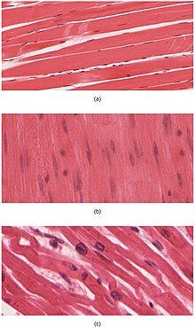
The body contains three types of muscle tissue: (a) skeletal muscle, (b) smooth muscle, and (c) cardiac muscle.

On the anterior and posterior views of the muscular system above, superficial muscles (those at the surface) are shown on the right side of the body while deep muscles (those underneath the superficial muscles) are shown on the left half of the body. For the legs, superficial muscles are shown in the anterior view while the posterior view shows both superficial and deep muscles.
There are three types of muscles—cardiac, skeletal, and smooth. Smooth muscles are used to control the flow of substances within the lumens of hollow organs, and are not consciously controlled. Skeletal and cardiac muscles have striations that are visible under a microscope due to the components within their cells. Only skeletal and smooth muscles are part of the musculoskeletal system and only the muscles can move the body. Cardiac muscles are found in the heart and are used only to circulate blood; like the smooth muscles, these muscles are not under conscious control. Skeletal muscles are attached to bones and arranged in opposing groups around joints.[8] Muscles are innervated, to communicate nervous energy to,[9] by nerves, which conduct electrical currents from the central nervous system and cause the muscles to contract.[10]
Contraction initiation[edit]
In mammals, when a muscle contracts, a series of reactions occur. Muscle contraction is stimulated by the motor neuron sending a message to the muscles from the somatic nervous system. Depolarization of the motor neuron results in neurotransmitters being released from the nerve terminal. The space between the nerve terminal and the muscle cell is called the neuromuscular junction. These neurotransmitters diffuse across the synapse and bind to specific receptor sites on the cell membrane of the muscle fiber. When enough receptors are stimulated, an action potential is generated and the permeability of the sarcolemma is altered. This process is known as initiation.[11]
Tendons[edit]
A tendon is a tough, flexible band of fibrous connective tissue that connects muscles to bones.[12] The extra-cellular connective tissue between muscle fibers binds to tendons at the distal and proximal ends, and the tendon binds to the periosteum of individual bones at the muscle’s origin and insertion. As muscles contract, tendons transmit the forces to the relatively rigid bones, pulling on them and causing movement. Tendons can stretch substantially, allowing them to function as springs during locomotion, thereby saving energy.
Joints, ligaments and bursae[edit]
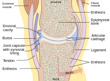
Human synovial joint composition
Joints are structures that connect individual bones and may allow bones to move against each other to cause movement. There are three divisions of joints, diarthroses which allow extensive mobility between two or more articular heads; amphiarthrosis, which is a joint that allows some movement, and false joints or synarthroses, joints that are immovable, that allow little or no movement and are predominantly fibrous. Synovial joints, joints that are not directly joined, are lubricated by a solution called synovial fluid that is produced by the synovial membranes. This fluid lowers the friction between the articular surfaces and is kept within an articular capsule, binding the joint with its taut tissue.[6]
Ligaments[edit]
A ligament is a small band of dense, white, fibrous elastic tissue.[6] Ligaments connect the ends of bones together in order to form a joint. Most ligaments limit dislocation, or prevent certain movements that may cause breaks. Since they are only elastic they increasingly lengthen when under pressure. When this occurs the ligament may be susceptible to break resulting in an unstable joint.
Ligaments may also restrict some actions: movements such as hyper extension and hyper flexion are restricted by ligaments to an extent. Also ligaments prevent certain directional movement.[13]
Bursae[edit]
A bursa is a small fluid-filled sac made of white fibrous tissue and lined with synovial membrane. Bursa may also be formed by a synovial membrane that extends outside of the joint capsule.[7] It provides a cushion between bones and tendons or muscles around a joint; bursa are filled with synovial fluid and are found around almost every major joint of the body.
Clinical significance[edit]

Because many other body systems, including the vascular, nervous, and integumentary systems, are interrelated, disorders of one of these systems may also affect the musculoskeletal system and complicate the diagnosis of the disorder’s origin. Diseases of the musculoskeletal system mostly encompass functional disorders or motion discrepancies; the level of impairment depends specifically on the problem and its severity. In a study of hospitalizations in the United States, the most common inpatient OR procedures in 2012 involved the musculoskeletal system: knee arthroplasty, laminectomy, hip replacement, and spinal fusion.[15]
Articular (of or pertaining to the joints)[16] disorders are the most common. However, also among the diagnoses are: primary muscular diseases, neurologic (related to the medical science that deals with the nervous system and disorders affecting it)[17] deficits, toxins, endocrine abnormalities, metabolic disorders, infectious diseases, blood and vascular disorders, and nutritional imbalances.
Disorders of muscles from another body system can bring about irregularities such as: impairment of ocular motion and control, respiratory dysfunction, and bladder malfunction. Complete paralysis, paresis, or ataxia may be caused by primary muscular dysfunctions of infectious or toxic origin; however, the primary disorder is usually related to the nervous system, with the muscular system acting as the effector organ, an organ capable of responding to a stimulus, especially a nerve impulse.[3]
One understated disorder that begins during pregnancy is pelvic girdle pain. It is complex, multi-factorial, and likely to be also represented by a series of sub-groups driven by pain varying from peripheral or central nervous system,[18] altered laxity/stiffness of muscles,[19] laxity to injury of tendinous/ligamentous structures[20]
to maladaptive body mechanics.[18]
See also[edit]
- Skeletal muscles of the human body
- Skeletal muscle
- Muscular system
References[edit]
- ^ Musculoskeletal+System at the US National Library of Medicine Medical Subject Headings (MeSH)
- ^ Mooar, Pekka (2007). «Muscles». Merck Manual. Retrieved 12 November 2008.
- ^ a b Kahn, Cynthia; Scott Line (2008). Musculoskeletal System Introduction: Introduction. NJ, USA: Merck & Co., Inc.
- ^ a b Applegate, Edith; Kent Van De Graaff. «The Skeletal System». Archived from the original on 3 June 2010. Retrieved 3 January 2009.
- ^ Engelbert, Phillis; Carol DeKane Nagel (2009). «The Human Body / How Many Bones Are In The Human Body?». U·X·L Science Fact Finder. eNotes.com, Inc. Retrieved 24 January 2009.
- ^ a b c Gary, Farr (25 June 2002). «The Musculoskeletal System». Archived from the original on 29 November 2014. Retrieved 18 November 2008.
- ^ a b «Skeletal System». 2001. Archived from the original on 25 February 2011. Retrieved 8 January 2009.
- ^ Mooar, Pekka (2007). «Muscles». The Merck Manuals Online Medical Library. Retrieved 16 November 2008.
- ^ «innervated». Dictionary.com. Dictionary.com, LLC. 2008. Retrieved 3 January 2009.
- ^ Bárány, Michael (2002). «SMOOTH MUSCLE». Retrieved 19 November 2008.
- ^ «The Mechanism of Muscle Contraction». Principles of Meat Science (4th Edition). Archived from the original on 17 February 2012. Retrieved 18 November 2008.
- ^ Jonathan, Cluett (2008). «Tendons». Retrieved 19 November 2008.
- ^ Bridwell, Keith. «Ligaments». Retrieved 16 March 2009.
- ^ «WHO Disease and injury country estimates». World Health Organization. 2009. Retrieved 11 November 2009.
- ^ Fingar KR, Stocks C, Weiss AJ, Steiner CA (December 2014). «Most Frequent Operating Room Procedures Performed in U.S. Hospitals, 2003–2012». HCUP Statistical Brief #186. Rockville, MD: Agency for Healthcare Research and Quality.
- ^ «articular». Random House Unabridged Dictionary. Random House, Inc. 2006. Retrieved 15 November 2008.
- ^ «neurologic». The American Heritage Dictionary of the English Language, Fourth Edition. Houghton Mifflin Company. 2006. Retrieved 15 November 2008.
- ^ a b Diagnosis and classification of pelvic girdle pain disorders— Part 1: A mechanism based approach within a bio psychosocial framework.
Manual Therapy, Volume 12, Issue 2, May 2007, PB. O’Sullivan and DJ Beales. - ^ Vleeming, Andry; Albert, Hanne B.; Östgaard, Hans Christian; Sturesson, Bengt; Stuge, Britt (June 2008). «European guidelines for the diagnosis and treatment of pelvic girdle pain». European Spine Journal. 17 (6): 794–819. doi:10.1007/s00586-008-0602-4. PMC 2518998. PMID 18259783.
- ^ Vleeming, Andry; de Vries, Haitze; Mens, Jan; van Wingerden, Jan-Paul (2002). «Possible role of the long dorsal sacroiliac ligament in women with peripartum pelvic pain». Acta Obstetricia et Gynecologica Scandinavica. 81 (5): 430–436. doi:10.1034/j.1600-0412.2002.810510.x. ISSN 0001-6349. PMID 12027817. S2CID 18323116.
From Wikipedia, the free encyclopedia
| Musculoskeletal system | |
|---|---|
|
Features of the human activity system from the 1911 Encyclopædia Britannica |
|
| Identifiers | |
| MeSH | D009141 |
| TA2 | 351 |
| FMA | 7482 |
| Anatomical terminology
[edit on Wikidata] |
The human musculoskeletal system (also known as the human locomotor system, and previously the activity system) is an organ system that gives humans the ability to move using their muscular and skeletal systems. The musculoskeletal system provides form, support, stability, and movement to the body.
It is made up of the bones of the skeleton, muscles, cartilage,[1] tendons, ligaments, joints, and other connective tissue that supports and binds tissues and organs together. The musculoskeletal system’s primary functions include supporting the body, allowing motion, and protecting vital organs.[2] The skeletal portion of the system serves as the main storage system for calcium and phosphorus and contains critical components of the hematopoietic system.[3]
This system describes how bones are connected to other bones and muscle fibers via connective tissue such as tendons and ligaments. The bones provide stability to the body. Muscles keep bones in place and also play a role in the movement of bones. To allow motion, different bones are connected by joints. Cartilage prevents the bone ends from rubbing directly onto each other. Muscles contract to move the bone attached at the joint.
There are, however, diseases and disorders that may adversely affect the function and overall effectiveness of the system. These diseases can be difficult to diagnose due to the close relation of the musculoskeletal system to other internal systems. The musculoskeletal system refers to the system having its muscles attached to an internal skeletal system and is necessary for humans to move to a more favorable position. Complex issues and injuries involving the musculoskeletal system are usually handled by a physiatrist (specialist in physical medicine and rehabilitation) or an orthopaedic surgeon.
Subsystems[edit]
Skeletal[edit]
The skeletal system serves many important functions; it provides the shape and form for the body, support and protection, allows bodily movement, produces blood for the body, and stores minerals.[4] The number of bones in the human skeletal system is a controversial topic. Humans are born with over 300 bones; however, many bones fuse together between birth and maturity. As a result, an average adult skeleton consists of 206 bones. The number of bones varies according to the method used to derive the count. While some consider certain structures to be a single bone with multiple parts, others may see it as a single part with multiple bones.[5] There are five general classifications of bones. These are long bones, short bones, flat bones, irregular bones, and sesamoid bones. The human skeleton is composed of both fused and individual bones supported by ligaments, tendons, muscles and cartilage. It is a complex structure with two distinct divisions; the axial skeleton, which includes the vertebral column, and the appendicular skeleton.[6]
Function[edit]
The skeletal system serves as a framework for tissues and organs to attach themselves to. This system acts as a protective structure for vital organs. Major examples of this are the brain being protected by the skull and the lungs being protected by the rib cage.
Located in long bones are two distinctions of bone marrow (yellow and red). The yellow marrow has fatty connective tissue and is found in the marrow cavity. During starvation, the body uses the fat in yellow marrow for energy.[7] The red marrow of some bones is an important site for blood cell production, approximately 2.6 million red blood cells per second in order to replace existing cells that have been destroyed by the liver.[4] Here all erythrocytes, platelets, and most leukocytes form in adults. From the red marrow, erythrocytes, platelets, and leukocytes migrate to the blood to do their special tasks.
Another function of bones is the storage of certain minerals. Calcium and phosphorus are among the main minerals being stored. The importance of this storage «device» helps to regulate mineral balance in the bloodstream. When the fluctuation of minerals is high, these minerals are stored in bone; when it is low it will be withdrawn from the bone.
Muscular[edit]
The body contains three types of muscle tissue: (a) skeletal muscle, (b) smooth muscle, and (c) cardiac muscle.
On the anterior and posterior views of the muscular system above, superficial muscles (those at the surface) are shown on the right side of the body while deep muscles (those underneath the superficial muscles) are shown on the left half of the body. For the legs, superficial muscles are shown in the anterior view while the posterior view shows both superficial and deep muscles.
There are three types of muscles—cardiac, skeletal, and smooth. Smooth muscles are used to control the flow of substances within the lumens of hollow organs, and are not consciously controlled. Skeletal and cardiac muscles have striations that are visible under a microscope due to the components within their cells. Only skeletal and smooth muscles are part of the musculoskeletal system and only the muscles can move the body. Cardiac muscles are found in the heart and are used only to circulate blood; like the smooth muscles, these muscles are not under conscious control. Skeletal muscles are attached to bones and arranged in opposing groups around joints.[8] Muscles are innervated, to communicate nervous energy to,[9] by nerves, which conduct electrical currents from the central nervous system and cause the muscles to contract.[10]
Contraction initiation[edit]
In mammals, when a muscle contracts, a series of reactions occur. Muscle contraction is stimulated by the motor neuron sending a message to the muscles from the somatic nervous system. Depolarization of the motor neuron results in neurotransmitters being released from the nerve terminal. The space between the nerve terminal and the muscle cell is called the neuromuscular junction. These neurotransmitters diffuse across the synapse and bind to specific receptor sites on the cell membrane of the muscle fiber. When enough receptors are stimulated, an action potential is generated and the permeability of the sarcolemma is altered. This process is known as initiation.[11]
Tendons[edit]
A tendon is a tough, flexible band of fibrous connective tissue that connects muscles to bones.[12] The extra-cellular connective tissue between muscle fibers binds to tendons at the distal and proximal ends, and the tendon binds to the periosteum of individual bones at the muscle’s origin and insertion. As muscles contract, tendons transmit the forces to the relatively rigid bones, pulling on them and causing movement. Tendons can stretch substantially, allowing them to function as springs during locomotion, thereby saving energy.
Joints, ligaments and bursae[edit]
Human synovial joint composition
Joints are structures that connect individual bones and may allow bones to move against each other to cause movement. There are three divisions of joints, diarthroses which allow extensive mobility between two or more articular heads; amphiarthrosis, which is a joint that allows some movement, and false joints or synarthroses, joints that are immovable, that allow little or no movement and are predominantly fibrous. Synovial joints, joints that are not directly joined, are lubricated by a solution called synovial fluid that is produced by the synovial membranes. This fluid lowers the friction between the articular surfaces and is kept within an articular capsule, binding the joint with its taut tissue.[6]
Ligaments[edit]
A ligament is a small band of dense, white, fibrous elastic tissue.[6] Ligaments connect the ends of bones together in order to form a joint. Most ligaments limit dislocation, or prevent certain movements that may cause breaks. Since they are only elastic they increasingly lengthen when under pressure. When this occurs the ligament may be susceptible to break resulting in an unstable joint.
Ligaments may also restrict some actions: movements such as hyper extension and hyper flexion are restricted by ligaments to an extent. Also ligaments prevent certain directional movement.[13]
Bursae[edit]
A bursa is a small fluid-filled sac made of white fibrous tissue and lined with synovial membrane. Bursa may also be formed by a synovial membrane that extends outside of the joint capsule.[7] It provides a cushion between bones and tendons or muscles around a joint; bursa are filled with synovial fluid and are found around almost every major joint of the body.
Clinical significance[edit]
Because many other body systems, including the vascular, nervous, and integumentary systems, are interrelated, disorders of one of these systems may also affect the musculoskeletal system and complicate the diagnosis of the disorder’s origin. Diseases of the musculoskeletal system mostly encompass functional disorders or motion discrepancies; the level of impairment depends specifically on the problem and its severity. In a study of hospitalizations in the United States, the most common inpatient OR procedures in 2012 involved the musculoskeletal system: knee arthroplasty, laminectomy, hip replacement, and spinal fusion.[15]
Articular (of or pertaining to the joints)[16] disorders are the most common. However, also among the diagnoses are: primary muscular diseases, neurologic (related to the medical science that deals with the nervous system and disorders affecting it)[17] deficits, toxins, endocrine abnormalities, metabolic disorders, infectious diseases, blood and vascular disorders, and nutritional imbalances.
Disorders of muscles from another body system can bring about irregularities such as: impairment of ocular motion and control, respiratory dysfunction, and bladder malfunction. Complete paralysis, paresis, or ataxia may be caused by primary muscular dysfunctions of infectious or toxic origin; however, the primary disorder is usually related to the nervous system, with the muscular system acting as the effector organ, an organ capable of responding to a stimulus, especially a nerve impulse.[3]
One understated disorder that begins during pregnancy is pelvic girdle pain. It is complex, multi-factorial, and likely to be also represented by a series of sub-groups driven by pain varying from peripheral or central nervous system,[18] altered laxity/stiffness of muscles,[19] laxity to injury of tendinous/ligamentous structures[20]
to maladaptive body mechanics.[18]
See also[edit]
- Skeletal muscles of the human body
- Skeletal muscle
- Muscular system
References[edit]
- ^ Musculoskeletal+System at the US National Library of Medicine Medical Subject Headings (MeSH)
- ^ Mooar, Pekka (2007). «Muscles». Merck Manual. Retrieved 12 November 2008.
- ^ a b Kahn, Cynthia; Scott Line (2008). Musculoskeletal System Introduction: Introduction. NJ, USA: Merck & Co., Inc.
- ^ a b Applegate, Edith; Kent Van De Graaff. «The Skeletal System». Archived from the original on 3 June 2010. Retrieved 3 January 2009.
- ^ Engelbert, Phillis; Carol DeKane Nagel (2009). «The Human Body / How Many Bones Are In The Human Body?». U·X·L Science Fact Finder. eNotes.com, Inc. Retrieved 24 January 2009.
- ^ a b c Gary, Farr (25 June 2002). «The Musculoskeletal System». Archived from the original on 29 November 2014. Retrieved 18 November 2008.
- ^ a b «Skeletal System». 2001. Archived from the original on 25 February 2011. Retrieved 8 January 2009.
- ^ Mooar, Pekka (2007). «Muscles». The Merck Manuals Online Medical Library. Retrieved 16 November 2008.
- ^ «innervated». Dictionary.com. Dictionary.com, LLC. 2008. Retrieved 3 January 2009.
- ^ Bárány, Michael (2002). «SMOOTH MUSCLE». Retrieved 19 November 2008.
- ^ «The Mechanism of Muscle Contraction». Principles of Meat Science (4th Edition). Archived from the original on 17 February 2012. Retrieved 18 November 2008.
- ^ Jonathan, Cluett (2008). «Tendons». Retrieved 19 November 2008.
- ^ Bridwell, Keith. «Ligaments». Retrieved 16 March 2009.
- ^ «WHO Disease and injury country estimates». World Health Organization. 2009. Retrieved 11 November 2009.
- ^ Fingar KR, Stocks C, Weiss AJ, Steiner CA (December 2014). «Most Frequent Operating Room Procedures Performed in U.S. Hospitals, 2003–2012». HCUP Statistical Brief #186. Rockville, MD: Agency for Healthcare Research and Quality.
- ^ «articular». Random House Unabridged Dictionary. Random House, Inc. 2006. Retrieved 15 November 2008.
- ^ «neurologic». The American Heritage Dictionary of the English Language, Fourth Edition. Houghton Mifflin Company. 2006. Retrieved 15 November 2008.
- ^ a b Diagnosis and classification of pelvic girdle pain disorders— Part 1: A mechanism based approach within a bio psychosocial framework.
Manual Therapy, Volume 12, Issue 2, May 2007, PB. O’Sullivan and DJ Beales. - ^ Vleeming, Andry; Albert, Hanne B.; Östgaard, Hans Christian; Sturesson, Bengt; Stuge, Britt (June 2008). «European guidelines for the diagnosis and treatment of pelvic girdle pain». European Spine Journal. 17 (6): 794–819. doi:10.1007/s00586-008-0602-4. PMC 2518998. PMID 18259783.
- ^ Vleeming, Andry; de Vries, Haitze; Mens, Jan; van Wingerden, Jan-Paul (2002). «Possible role of the long dorsal sacroiliac ligament in women with peripartum pelvic pain». Acta Obstetricia et Gynecologica Scandinavica. 81 (5): 430–436. doi:10.1034/j.1600-0412.2002.810510.x. ISSN 0001-6349. PMID 12027817. S2CID 18323116.
Предложения со словосочетанием «опорно-двигательная система»

Кости нужно массировать, разминать, чтобы костный мозг получал достаточно питания. Наша опорно-двигательная система должна быть живой.
Прежде чем вы перейдёте собственно к рассмотрению различных упражнений, вам полезно будет подробнее узнать о том, что же представляют собой мышечная, дыхательная и опорно-двигательная системы человека.
Опорно-двигательная система людей этих профессий испытывает колоссальные физические нагрузки и крайне нуждается в реабилитации.
Важную роль в борьбе с недугами и преодолении их играет наша опорно-двигательная система.
В большей степени страдают сердечно-сосудистая, дыхательная и опорно-двигательная системы, печень и кожа.

Привет! Меня зовут Лампобот, я компьютерная программа, которая помогает делать
Карту слов. Я отлично
умею считать, но пока плохо понимаю, как устроен ваш мир. Помоги мне разобраться!
Спасибо! Я стал чуточку лучше понимать мир эмоций.
Вопрос: автокефалия — это что-то нейтральное, положительное или отрицательное?
Внутренние органы, конечности (ноги и руки, стопы и ладони, пальцы рук и ног), чувства (слух, зрение, вкус, запах, тактильные ощущения) и опорно-двигательная система (осанка и ориентация) – всё это находится в непрерывном двустороннем взаимодействии с островковой долей мозга, центральной частью мозга, в которой физиологический опыт соединяется с мыслями и чувствами – и наоборот.
К тому же мы уже его многому научили: опорно-двигательная система уже работает на сто процентов, он пользуется транслейтером.
Поскольку опорно-двигательная система – это наш каркас, опора и основа, то её разбалансированность провоцирует болезни других систем и отдельных органов, а также снижение подвижности и ухудшение общего состояния организма.
За время, проведённое на больничной койке, у меня атрофировалась опорно-двигательная система.
Опорно-двигательная система человека образована скелетом и мышцами и представляет собой автоматизированную систему, в которой всё должно быть чётко отрегулировано.
Вообще, каждый человек должен знать, как работает опорно-двигательная система, как наш скелет двигается.
А при том, что опорно-двигательная система представляет собой единое целое, а не простую совокупность отдельных мышц и суставов.
У него была гармонично развита опорно-двигательная система, хорошая сердечно-сосудистая система.
Опорно-двигательная система — это тот каркас, который позволяет нам изменять своё положение в пространстве и формирует наш внешний облик.
Опорно-двигательная система защищает наши важнейшие органы и системы от неблагоприятных воздействий внешней среды.
Первые шаги давались сложно, опорно-двигательная система барахлила.
Мужчине нет сорока, а он стал замечать, что возникли проблемы сразу по нескольким физиологическим фронтам: желудочно-кишечный тракт и опорно-двигательная система перестали работать идеально, а ко всему прочему стала вдруг подводить память.
– Записывай, – резко бросил он, с неприязнью глядя на меня, – Опорно-двигательная система отказывает, речь не проявляется.
Его опорно-двигательная система непрерывно осуществляет статическую работу по адекватному противодействию силе, с которой земля притягивает тело к себе.
Ассоциации к слову «система»
Синонимы к слову «система»
Цитаты из русской классики со словосочетанием «опорно-двигательная система»
- Он жил безвыездно в Москве, в Пименовском переулке, близ Малой Дмитровки, в собственном доме, над крышей которого высилась целая система громоотводов, но они, впрочем, не рассеивали его опасения, и старик во время грозы скрывался на погребице.
- Но до того человек пристрастен к системе и к отвлеченному выводу, что готов умышленно исказить правду, готов видом не видать и слыхом не слыхать, только чтоб оправдать свою логику.
- Такой системы Ребер и держался в первых двух состязаниях, из которых одно осталось за Арбузовым, а другое за ним.
- (все
цитаты из русской классики)
Сочетаемость слова «система»
- нервная система
солнечная система
иммунная система - система управления
система образования
система безопасности - работа нервной системы
создание системы
развитие системы - система работает
система рухнет
система существует - создать систему
войти в систему
иметь систему - (полная таблица сочетаемости)
Значение слова «система»
-
СИСТЕ́МА, -ы, ж. 1. Множество элементов, находящихся в отношениях и связях друг с другом и образующих определенную целостность, единство. (Малый академический словарь, МАС)
Все значения слова СИСТЕМА
Афоризмы русских писателей со словом «система»
- Создать язык невозможно, ибо его творит народ; филологи только открывают его законы и приводят их в систему, а писатели только творят на нем сообразно с сими законами.
- Свое собственное, вольное и свободное хотенье, свой собственный, хотя бы и самый дикий каприз, своя фантазия, раздраженная иногда хоть бы даже до сумасшествия, — вот это-то все и есть та самая, самая выгодная выгода, которая ни под какую классификацию не подходит и от которой все теории и системы постоянно разлетаются к черту. И с чего это взяли все эти мудрецы, что человеку надо какого-то нормального, какого-то добродетельного хотения? С чего это непременно вообразили они, что человеку надо непременно благоразумно выгодного хотенья? Человеку надо — одного самостоятельного хотенья, чего бы эта самостоятельность ни стоила и к чему бы ни привела.
- Англичанин молчалив, равнодушен, говорит, как читает, не обнаруживая никогда быстрых душевных стремлений, которые потрясают электрически всю нашу физическую систему.
- (все афоризмы русских писателей)
Отправить комментарий
Дополнительно
Морфемы слова опорно-двигательный
Разбор слова «опорно-двигательный» по составу: слово опорно-двигательный имеет приставка «о», корень «пор», суффикс «н», соединительную гласную «о», корень «двиг», суффикс «а», суффикс «тельн», окончание «ый»
опорно-двигательный
Как пишется слово опорно-двигательный?
Правильно пишется: опорно-двигательный
Разбор слова «опорно-двигательный»
| приставка | о |
| корень | пор |
| суффикс | н |
| соединительная гласная |
о |
| корень | двиг |
| суффикс | а |
| суффикс | тельн |
| окончание | ый |
Примеры со словом опорно-двигательный
Очень уязвим опорно-двигательный аппарат.
Может, предательски отказавший опорно-двигательный аппарат.
Узнайте как пишется слово «засевать»
39% ошибается при написании этого слова, проверьте свои знания
![]()
Слова русского языка,
поиск и разбор слов онлайн
опорно-двигательный
Правильно слово пишется: опо́рно-дви́гательный
Сложное слово, состоящее из 2 частей.
- опорно
- Ударение падает на 2-й слог с буквой о.
Всего в слове 6 букв, 3 гласных, 3 согласных, 3 слога.
Гласные: о, о, о;
Согласные: п, р, н. - двигательный
- Ударение падает на 1-й слог с буквой и.
Всего в слове 12 букв, 4 гласных, 7 согласных, 4 слога.
Гласные: и, а, е, ы;
Согласные: д, в, г, т, л, н, й;
1 буква не обозначает звука.
Номера букв в слове
Номера букв в слове «опорно-двигательный» в прямом и обратном порядке:
- 18
о
1 - 17
п
2 - 16
о
3 - 15
р
4 - 14
н
5 - 13
о
6 -
—
- 12
д
7 - 11
в
8 - 10
и
9 - 9
г
10 - 8
а
11 - 7
т
12 - 6
е
13 - 5
л
14 - 4
ь
15 - 3
н
16 - 2
ы
17 - 1
й
18
Слово «опорно-двигательный» состоит из 18-ти букв и 1-го дефиса.
Разбор по составу
Разбор по составу (морфемный разбор) слова опорно-двигательный делается следующим образом:
опорно—двигательный
Морфемы слова: о —приставка, пор, двиг — корни, н, о, а, тельн — суффиксы, ый — окончание, опорно-двигательн — основы.