РусскийПравить
Морфологические и синтаксические свойстваПравить
| падеж | ед. ч. | мн. ч. |
|---|---|---|
| Им. | тахикарди́я | тахикарди́и |
| Р. | тахикарди́и | тахикарди́й |
| Д. | тахикарди́и | тахикарди́ям |
| В. | тахикарди́ю | тахикарди́и |
| Тв. | тахикарди́ей тахикарди́ею |
тахикарди́ями |
| Пр. | тахикарди́и | тахикарди́ях |
та—хи—кар—ди́·я
Существительное, неодушевлённое, женский род, 1-е склонение (тип склонения 7a по классификации А. А. Зализняка).
Корень: -тахи-; корень: -кард-; суффикс: -иj; окончание: -я [Тихонов, 1996].
ПроизношениеПравить
- МФА: [təxʲɪkɐrˈdʲiɪ̯ə]
Семантические свойстваПравить
ЗначениеПравить
- мед. увеличение частоты сердечных сокращений до 100-180 в минуту, возникающее при физическом и нервных напряжениях, заболеваниях сердечно-сосудистой и нервной систем, болезнях желёз внутренней секреции и др. ◆ Заболевания щитовидной железы обычно приводят к постоянной тахикардии, сопровождающейся к тому же дрожанием пальцев рук, век, повышенной потливостью. «Большие проблемы маленькой железы», 1999 г. // «Здоровье» [НКРЯ]
СинонимыПравить
АнтонимыПравить
- брадикардия
ГиперонимыПравить
- учащение
ГипонимыПравить
Родственные словаПравить
| Ближайшее родство | |
ЭтимологияПравить
Происходит от лат. tachycardia «тахикардия», составленного из др.-греч. ταχύς «быстрый», далее из неустановленной формы, + др.-греч. καρδία «сердце», далее из праиндоевр. *kerd- «сердце; сердцевина, середина».
Фразеологизмы и устойчивые сочетанияПравить
ПереводПравить
| Список переводов | |
|
БиблиографияПравить
БашкирскийПравить
Морфологические и синтаксические свойстваПравить
тахикардия
Существительное.
Корень: —.
ПроизношениеПравить
Семантические свойстваПравить
ЗначениеПравить
- мед. тахикардия ◆ Отсутствует пример употребления (см. рекомендации).
СинонимыПравить
АнтонимыПравить
ГиперонимыПравить
ГипонимыПравить
Родственные словаПравить
| Ближайшее родство | |
ЭтимологияПравить
От лат. tachycardia «тахикардия», составленного из др.-греч. ταχύς «быстрый», далее из неустановленной формы, + др.-греч. καρδία «сердце», далее из праиндоевр. *kerd- «сердце; сердцевина, середина».
Фразеологизмы и устойчивые сочетанияПравить
БиблиографияПравить
БолгарскийПравить
Морфологические и синтаксические свойстваПравить
тахикардия
Существительное, женский род.
Корень: —.
ПроизношениеПравить
Семантические свойстваПравить
ЗначениеПравить
- мед. тахикардия ◆ Отсутствует пример употребления (см. рекомендации).
СинонимыПравить
АнтонимыПравить
ГиперонимыПравить
ГипонимыПравить
Родственные словаПравить
| Ближайшее родство | |
ЭтимологияПравить
От лат. tachycardia «тахикардия», составленного из др.-греч. ταχύς «быстрый», далее из неустановленной формы, + др.-греч. καρδία «сердце», далее из праиндоевр. *kerd- «сердце; сердцевина, середина».
Фразеологизмы и устойчивые сочетанияПравить
БиблиографияПравить
КазахскийПравить
Морфологические и синтаксические свойстваПравить
тахикардия
Существительное.
Корень: —.
ПроизношениеПравить
Семантические свойстваПравить
ЗначениеПравить
- мед. тахикардия ◆ Отсутствует пример употребления (см. рекомендации).
СинонимыПравить
АнтонимыПравить
ГиперонимыПравить
ГипонимыПравить
Родственные словаПравить
| Ближайшее родство | |
ЭтимологияПравить
От лат. tachycardia «тахикардия», составленного из др.-греч. ταχύς «быстрый», далее из неустановленной формы, + др.-греч. καρδία «сердце», далее из праиндоевр. *kerd- «сердце; сердцевина, середина».
Фразеологизмы и устойчивые сочетанияПравить
БиблиографияПравить
ТаджикскийПравить
Морфологические и синтаксические свойстваПравить
тахикардия
Существительное.
Корень: —.
ПроизношениеПравить
Семантические свойстваПравить
ЗначениеПравить
- мед. тахикардия ◆ Отсутствует пример употребления (см. рекомендации).
СинонимыПравить
АнтонимыПравить
ГиперонимыПравить
ГипонимыПравить
Родственные словаПравить
| Ближайшее родство | |
ЭтимологияПравить
От лат. tachycardia «тахикардия», составленного из др.-греч. ταχύς «быстрый», далее из неустановленной формы, + др.-греч. καρδία «сердце», далее из праиндоевр. *kerd- «сердце; сердцевина, середина».
Фразеологизмы и устойчивые сочетанияПравить
БиблиографияПравить
ТатарскийПравить
Морфологические и синтаксические свойстваПравить
тахикардия
Существительное.
Корень: —.
ПроизношениеПравить
Семантические свойстваПравить
ЗначениеПравить
- мед. тахикардия ◆ Отсутствует пример употребления (см. рекомендации).
СинонимыПравить
АнтонимыПравить
ГиперонимыПравить
ГипонимыПравить
Родственные словаПравить
| Ближайшее родство | |
ЭтимологияПравить
От лат. tachycardia «тахикардия», составленного из др.-греч. ταχύς «быстрый», далее из неустановленной формы, + др.-греч. καρδία «сердце», далее из праиндоевр. *kerd- «сердце; сердцевина, середина».
Фразеологизмы и устойчивые сочетанияПравить
БиблиографияПравить
From Wikipedia, the free encyclopedia
| Tachycardia | |
|---|---|
| Other names | Tachyarrhythmia |
 |
|
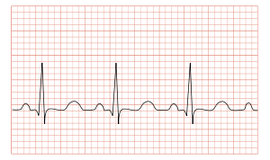
| ECG showing sinus tachycardia with a rate of about 100 beats per minute | |
| Pronunciation |
|
| Specialty | Cardiology |
| Differential diagnosis |
|
Tachycardia, also called tachyarrhythmia, is a heart rate that exceeds the normal resting rate.[1] In general, a resting heart rate over 100 beats per minute is accepted as tachycardia in adults.[1] Heart rates above the resting rate may be normal (such as with exercise) or abnormal (such as with electrical problems within the heart).
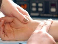
Auscultation of a 14 year old female’s heart during an episode of tachyarrhythmia.
Complications[edit]
Tachycardia can lead to fainting.[2]
When the rate of blood flow becomes too rapid, or fast blood flow passes on damaged endothelium, it increases the friction within vessels resulting in turbulence and other disturbances.[3] According to the Virchow’s triad, this is one of the three conditions that can lead to thrombosis (i.e., blood clots within vessels).[citation needed]
Causes[edit]
Some causes of tachycardia include:[4]
- Adrenergic storm
- Anaemia
- Anxiety
- Atrial fibrillation
- Atrial flutter
- Atrial tachycardia
- Atrioventricular reentrant tachycardia
- AV nodal reentrant tachycardia
- Brugada syndrome
- Circulatory shock and its various causes (obstructive shock, cardiogenic shock, hypovolemic shock, distributive shock)
- Dysautonomia
- Exercise
- Fear
- Hypoglycemia
- Hypovolemia
- Hyperthyroidism
- Hyperventilation
- Inappropriate sinus tachycardia
- Junctional tachycardia
- Metabolic myopathy
- Multifocal atrial tachycardia
- Pacemaker mediated
- Pain
- Panic attack
- Pheochromocytoma
- Sinus tachycardia
- Sleep deprivation[5]
- Supraventricular tachycardia
- Ventricular tachycardia
- Wolff–Parkinson–White syndrome
Drug related:
- Alcohol (Ethanol) intoxication
- Stimulants
- Cannabis
- Drug withdrawal
- Tricyclic antidepressants
- Nefopam
- Opioids (rare)
Diagnosis[edit]
The upper threshold of a normal human resting heart rate is based on age. Cutoff values for tachycardia in different age groups are fairly well standardized; typical cutoffs are listed below:[6]
- 1–2 days: Tachycardia >159 beats per minute (bpm)
- 3–6 days: Tachycardia >166 bpm
- 1–3 weeks: Tachycardia >182 bpm
- 1–2 months: Tachycardia >179 bpm
- 3–5 months: Tachycardia >186 bpm
- 6–11 months: Tachycardia >169 bpm
- 1–2 years: Tachycardia >151 bpm
- 3–4 years: Tachycardia >137 bpm
- 5–7 years: Tachycardia >133 bpm
- 8–11 years: Tachycardia >130 bpm
- 12–15 years: Tachycardia >119 bpm
- >15 years – adult: Tachycardia >100 bpm
Heart rate is considered in the context of the prevailing clinical picture. When the heart beats excessively or rapidly, the heart pumps less efficiently and provides less blood flow to the rest of the body, including the heart itself. The increased heart rate also leads to increased work and oxygen demand by the heart, which can lead to rate related ischemia.[7]
Differential diagnosis[edit]
An electrocardiogram (ECG) is used to classify the type of tachycardia. They may be classified into narrow and wide complex based on the QRS complex.[8] Equal or less than 0.1s for narrow complex.[9] Presented in order of most to least common, they are:[8]
- Narrow complex
- Sinus tachycardia, which originates from the sino-atrial (SA) node, near the base of the superior vena cava
- Atrial fibrillation
- Atrial flutter
- AV nodal reentrant tachycardia
- Accessory pathway mediated tachycardia
- Atrial tachycardia
- Multifocal atrial tachycardia
- Cardiac Tamponade
- Junctional tachycardia (rare in adults)
- Wide complex
- Ventricular tachycardia, any tachycardia that originates in the ventricles
- Any narrow complex tachycardia combined with a problem with the conduction system of the heart, often termed «supraventricular tachycardia with aberrancy»
- A narrow complex tachycardia with an accessory conduction pathway, often termed «supraventricular tachycardia with pre-excitation» (e.g. Wolff–Parkinson–White syndrome)
- Pacemaker-tracked or pacemaker-mediated tachycardia
Tachycardias may be classified as either narrow complex tachycardias (supraventricular tachycardias) or wide complex tachycardias. Narrow and wide refer to the width of the QRS complex on the ECG. Narrow complex tachycardias tend to originate in the atria, while wide complex tachycardias tend to originate in the ventricles. Tachycardias can be further classified as either regular or irregular.[citation needed]
Sinus[edit]
The body has several feedback mechanisms to maintain adequate blood flow and blood pressure. If blood pressure decreases, the heart beats faster in an attempt to raise it. This is called reflex tachycardia. This can happen in response to a decrease in blood volume (through dehydration or bleeding), or an unexpected change in blood flow. The most common cause of the latter is orthostatic hypotension (also called postural hypotension). Fever, hyperventilation, diarrhea and severe infections can also cause tachycardia, primarily due to increase in metabolic demands.[citation needed]
Upon exertion, sinus tachycardia can also be seen in some inborn errors of metabolism that result in metabolic myopathies, such as McArdle’s disease (GSD-V).[10][11] Metabolic myopathies interfere with the muscle’s ability to create energy. This energy shortage in muscle cells causes an inappropriate rapid heart rate in response to exercise. The heart tries to compensate for the energy shortage by increasing heart rate to maximize delivery of oxygen and other blood borne fuels to the muscle cells.[10]
«In McArdle’s, our heart rate tends to increase in what is called an ‘inappropriate’ response. That is, after the start of exercise it increases much more quickly than would be expected in someone unaffected by McArdle’s.»[12] As skeletal muscle relies predominantly on glycogenolysis for the first few minutes as it transitions from rest to activity, as well as throughout high-intensity aerobic activity and all anaerobic activity, individuals with GSD-V experience during exercise: sinus tachycardia, tachypnea, muscle fatigue and pain, during the aforementioned activities and time frames.[10][11] Those with GSD-V also experience «second wind,» after approximately 6-10 minutes of light-moderate aerobic activity, such as walking without an incline, where the heart rate drops and symptoms of exercise intolerance improve.[10][11][12]
An increase in sympathetic nervous system stimulation causes the heart rate to increase, both by the direct action of sympathetic nerve fibers on the heart and by causing the endocrine system to release hormones such as epinephrine (adrenaline), which have a similar effect. Increased sympathetic stimulation is usually due to physical or psychological stress. This is the basis for the so-called fight-or-flight response, but such stimulation can also be induced by stimulants such as ephedrine, amphetamines or cocaine. Certain endocrine disorders such as pheochromocytoma can also cause epinephrine release and can result in tachycardia independent of nervous system stimulation. Hyperthyroidism can also cause tachycardia.[13] The upper limit of normal rate for sinus tachycardia is thought to be 220 bpm minus age.[citation needed]
Inappropriate sinus tachycardia[edit]
Inappropriate sinus tachycardia (IST) is a rare but benign type of cardiac arrhythmia that may be caused by a structural abnormality in the sinus node. It can occur in seemingly healthy individuals with no history of cardiovascular disease. Other causes may include autonomic nervous system deficits, autoimmune response, or drug interactions. Although symptoms might be distressing, treatment is not generally needed.[citation needed]
Ventricular[edit]
Ventricular tachycardia (VT or V-tach) is a potentially life-threatening cardiac arrhythmia that originates in the ventricles. It is usually a regular, wide complex tachycardia with a rate between 120 and 250 beats per minute. A medically significant subvariant of ventricular tachycardia is called torsades de pointes (literally meaning «twisting of the points», due to its appearance on an EKG), which tends to result from a long QT interval.[14]
Both of these rhythms normally last for only a few seconds to minutes (paroxysmal tachycardia), but if VT persists it is extremely dangerous, often leading to ventricular fibrillation.[citation needed]
Supraventricular[edit]
This is a type of tachycardia that originates from above the ventricles, such as the atria. It is sometimes known as paroxysmal atrial tachycardia (PAT). Several types of supraventricular tachycardia are known to exist.[15]
Atrial fibrillation[edit]
Atrial fibrillation is one of the most common cardiac arrhythmias. In general, it is an irregular, narrow complex rhythm. However, it may show wide QRS complexes on the ECG if a bundle branch block is present. At high rates, the QRS complex may also become wide due to the Ashman phenomenon. It may be difficult to determine the rhythm’s regularity when the rate exceeds 150 beats per minute. Depending on the patient’s health and other variables such as medications taken for rate control, atrial fibrillation may cause heart rates that span from 50 to 250 beats per minute (or even higher if an accessory pathway is present). However, new-onset atrial fibrillation tends to present with rates between 100 and 150 beats per minute.[16]
AV nodal reentrant tachycardia[edit]
AV nodal reentrant tachycardia (AVNRT) is the most common reentrant tachycardia. It is a regular narrow complex tachycardia that usually responds well to the Valsalva maneuver or the drug adenosine. However, unstable patients sometimes require synchronized cardioversion. Definitive care may include catheter ablation.[citation needed]
AV reentrant tachycardia[edit]
AV reentrant tachycardia (AVRT) requires an accessory pathway for its maintenance. AVRT may involve orthodromic conduction (where the impulse travels down the AV node to the ventricles and back up to the atria through the accessory pathway) or antidromic conduction (which the impulse travels down the accessory pathway and back up to the atria through the AV node). Orthodromic conduction usually results in a narrow complex tachycardia, and antidromic conduction usually results in a wide complex tachycardia that often mimics ventricular tachycardia. Most antiarrhythmics are contraindicated in the emergency treatment of AVRT, because they may paradoxically increase conduction across the accessory pathway.[citation needed]
Junctional tachycardia[edit]
Junctional tachycardia is an automatic tachycardia originating in the AV junction. It tends to be a regular, narrow complex tachycardia and may be a sign of digitalis toxicity.[citation needed]
Management[edit]
The management of tachycardia depends on its type (wide complex versus narrow complex), whether or not the person is stable or unstable, and whether the instability is due to the tachycardia.[8] Unstable means that either important organ functions are affected or cardiac arrest is about to occur.[8]
Unstable[edit]
In those that are unstable with a narrow complex tachycardia, intravenous adenosine may be attempted.[8] In all others immediate cardioversion is recommended.[8]
Terminology[edit]
The word tachycardia came to English from New Latin as a neoclassical compound built from the combining forms tachy- + -cardia, which are from the Greek ταχύς tachys, «quick, rapid» and καρδία, kardia, «heart». As a matter both of usage choices in the medical literature and of idiom in natural language, the words tachycardia and tachyarrhythmia are usually used interchangeably, or loosely enough that precise differentiation is not explicit. Some careful writers have tried to maintain a logical differentiation between them, which is reflected in major medical dictionaries[17][18][19] and major general dictionaries.[20][21][22] The distinction is that tachycardia be reserved for the rapid heart rate itself, regardless of cause, physiologic or pathologic (that is, from healthy response to exercise or from cardiac arrhythmia), and that tachyarrhythmia be reserved for the pathologic form (that is, an arrhythmia of the rapid rate type). This is why five of the previously referenced dictionaries do not enter cross-references indicating synonymy between their entries for the two words (as they do elsewhere whenever synonymy is meant), and it is why one of them explicitly specifies that the two words not be confused.[19] But the prescription will probably never be successfully imposed on general usage, not only because much of the existing medical literature ignores it even when the words stand alone but also because the terms for specific types of arrhythmia (standard collocations of adjectives and noun) are deeply established idiomatically with the tachycardia version as the more commonly used version. Thus SVT is called supraventricular tachycardia more than twice as often as it is called supraventricular tachyarrhythmia; moreover, those two terms are always completely synonymous—in natural language there is no such term as «healthy/physiologic supraventricular tachycardia». The same themes are also true of AVRT and AVNRT. Thus this pair is an example of when a particular prescription (which may have been tenable 50 or 100 years earlier) can no longer be invariably enforced without violating idiom. But the power to differentiate in an idiomatic way is not lost, regardless, because when the specification of physiologic tachycardia is needed, that phrase aptly conveys it.[citation needed]
See also[edit]
- Metabolic myopathies
- Postural orthostatic tachycardia syndrome
References[edit]
- ^ a b Awtry, Eric H.; Jeon, Cathy; Ware, Molly G. (2006). Blueprints cardiology (2nd ed.). Malden, Mass.: Blackwell. p. 93. ISBN 9781405104647.
- ^ «Passing Out (Syncope) Caused by Arrhythmias». Archived from the original on 2020-06-13. Retrieved 2020-04-13.
- ^ Kushner, Abigail; West, William P.; Pillarisetty, Leela Sharath (2020), «Virchow Triad», StatPearls, Treasure Island (FL): StatPearls Publishing, PMID 30969519, retrieved 2020-06-18
- ^ «Supraventricular Tachycardias». The Lecturio Medical Concept Library. 9 September 2020. Retrieved 2 July 2021.
- ^ Rangaraj VR, Knutson KL (February 2016). «Association between sleep deficiency and cardiometabolic disease: implications for health disparities». Sleep Med. 18: 19–35. doi:10.1016/j.sleep.2015.02.535. PMC 4758899. PMID 26431758.
- ^ Custer JW, Rau RE, eds. Johns Hopkins: The Harriet Lane Handbook. 18th ed. Philadelphia, PA: Mosby Elsevier Inc; 2008. Data also available through eMedicine: Pediatrics, Tachycardia.
- ^ Harrison’s Principles of Internal Medicine, 17th Edition
- ^ a b c d e f Neumar RW, Otto CW, Link MS, et al. (November 2010). «Part 8: adult advanced cardiovascular life support: 2010 American Heart Association Guidelines for Cardiopulmonary Resuscitation and Emergency Cardiovascular Care». Circulation. 122 (18 Suppl 3): S729–67. doi:10.1161/CIRCULATIONAHA.110.970988. PMID 20956224.
- ^ Pieper, Stephen J.; Stanton, Marshall S. (April 1995). «Narrow QRS Complex Tachycardias». Mayo Clinic Proceedings. 70 (4): 371–375. doi:10.4065/70.4.371. PMID 7898144.
- ^ a b c d Lucia A, Martinuzzi A, Nogales-Gadea G, Quinlivan R, Reason S; International Association for Muscle Glycogen Storage Disease study group. Clinical practice guidelines for glycogen storage disease V & VII (McArdle disease and Tarui disease) from an international study group. Neuromuscul Disord. 2021 Dec;31(12):1296-1310. doi: 10.1016/j.nmd.2021.10.006. Epub 2021 Oct 28. Erratum in: Neuromuscul Disord. 2022 Feb 6;: PMID: 34848128.
- ^ a b c Scalco RS, Chatfield S, Godfrey R, Pattni J, Ellerton C, Beggs A, Brady S, Wakelin A, Holton JL, Quinlivan R. From exercise intolerance to functional improvement: the second wind phenomenon in the identification of McArdle disease. Arq Neuropsiquiatr. 2014 Jul;72(7):538-41. doi: 10.1590/0004-282×20140062. PMID: 25054987.
- ^ a b Wakelin, Andrew (2017). Living With McArdle Disease (PDF). IAMGSD (International Assoc. of Muscle Glycogen Diseases). p. 15.
- ^ Barker RL, Burton JR, Zieve, PD eds. Principles of Ambulatory Medicine. Sixth Edition. Philadelphia, PA: Lippinocott, Wilkins & Williams 2003
- ^ MERCK Manual Profesional Edition RET. April 19, 2019 18:37 CST. https://www.merckmanuals.com/professional/cardiovascular-disorders/arrhythmias-and-conduction-disorders/long-qt-syndrome-and-torsades-de-pointes-ventricular-tachycardia
- ^ «Types of Arrhythmia». NHLBI. July 1, 2011. Archived from the original on June 7, 2015.
- ^ «Atrial Fibrillation». The Lecturio Medical Concept Library. 11 August 2020. Retrieved 3 July 2021.
- ^ Elsevier, Dorland’s Illustrated Medical Dictionary, Elsevier.
- ^ Merriam-Webster, Merriam-Webster’s Medical Dictionary, Merriam-Webster.
- ^ a b Wolters Kluwer, Stedman’s Medical Dictionary, Wolters Kluwer.
- ^ Houghton Mifflin Harcourt, The American Heritage Dictionary of the English Language, Houghton Mifflin Harcourt.
- ^ Merriam-Webster, Merriam-Webster’s Collegiate Dictionary, Merriam-Webster, archived from the original on 2020-10-10, retrieved 2017-07-22.
- ^ Merriam-Webster, Merriam-Webster’s Unabridged Dictionary, Merriam-Webster, archived from the original on 2020-05-25, retrieved 2017-07-22.
External links[edit]
From Wikipedia, the free encyclopedia
| Tachycardia | |
|---|---|
| Other names | Tachyarrhythmia |
 |
|
| ECG showing sinus tachycardia with a rate of about 100 beats per minute | |
| Pronunciation |
|
| Specialty | Cardiology |
| Differential diagnosis |
|
Tachycardia, also called tachyarrhythmia, is a heart rate that exceeds the normal resting rate.[1] In general, a resting heart rate over 100 beats per minute is accepted as tachycardia in adults.[1] Heart rates above the resting rate may be normal (such as with exercise) or abnormal (such as with electrical problems within the heart).
Auscultation of a 14 year old female’s heart during an episode of tachyarrhythmia.
Complications[edit]
Tachycardia can lead to fainting.[2]
When the rate of blood flow becomes too rapid, or fast blood flow passes on damaged endothelium, it increases the friction within vessels resulting in turbulence and other disturbances.[3] According to the Virchow’s triad, this is one of the three conditions that can lead to thrombosis (i.e., blood clots within vessels).[citation needed]
Causes[edit]
Some causes of tachycardia include:[4]
- Adrenergic storm
- Anaemia
- Anxiety
- Atrial fibrillation
- Atrial flutter
- Atrial tachycardia
- Atrioventricular reentrant tachycardia
- AV nodal reentrant tachycardia
- Brugada syndrome
- Circulatory shock and its various causes (obstructive shock, cardiogenic shock, hypovolemic shock, distributive shock)
- Dysautonomia
- Exercise
- Fear
- Hypoglycemia
- Hypovolemia
- Hyperthyroidism
- Hyperventilation
- Inappropriate sinus tachycardia
- Junctional tachycardia
- Metabolic myopathy
- Multifocal atrial tachycardia
- Pacemaker mediated
- Pain
- Panic attack
- Pheochromocytoma
- Sinus tachycardia
- Sleep deprivation[5]
- Supraventricular tachycardia
- Ventricular tachycardia
- Wolff–Parkinson–White syndrome
Drug related:
- Alcohol (Ethanol) intoxication
- Stimulants
- Cannabis
- Drug withdrawal
- Tricyclic antidepressants
- Nefopam
- Opioids (rare)
Diagnosis[edit]
The upper threshold of a normal human resting heart rate is based on age. Cutoff values for tachycardia in different age groups are fairly well standardized; typical cutoffs are listed below:[6]
- 1–2 days: Tachycardia >159 beats per minute (bpm)
- 3–6 days: Tachycardia >166 bpm
- 1–3 weeks: Tachycardia >182 bpm
- 1–2 months: Tachycardia >179 bpm
- 3–5 months: Tachycardia >186 bpm
- 6–11 months: Tachycardia >169 bpm
- 1–2 years: Tachycardia >151 bpm
- 3–4 years: Tachycardia >137 bpm
- 5–7 years: Tachycardia >133 bpm
- 8–11 years: Tachycardia >130 bpm
- 12–15 years: Tachycardia >119 bpm
- >15 years – adult: Tachycardia >100 bpm
Heart rate is considered in the context of the prevailing clinical picture. When the heart beats excessively or rapidly, the heart pumps less efficiently and provides less blood flow to the rest of the body, including the heart itself. The increased heart rate also leads to increased work and oxygen demand by the heart, which can lead to rate related ischemia.[7]
Differential diagnosis[edit]
An electrocardiogram (ECG) is used to classify the type of tachycardia. They may be classified into narrow and wide complex based on the QRS complex.[8] Equal or less than 0.1s for narrow complex.[9] Presented in order of most to least common, they are:[8]
- Narrow complex
- Sinus tachycardia, which originates from the sino-atrial (SA) node, near the base of the superior vena cava
- Atrial fibrillation
- Atrial flutter
- AV nodal reentrant tachycardia
- Accessory pathway mediated tachycardia
- Atrial tachycardia
- Multifocal atrial tachycardia
- Cardiac Tamponade
- Junctional tachycardia (rare in adults)
- Wide complex
- Ventricular tachycardia, any tachycardia that originates in the ventricles
- Any narrow complex tachycardia combined with a problem with the conduction system of the heart, often termed «supraventricular tachycardia with aberrancy»
- A narrow complex tachycardia with an accessory conduction pathway, often termed «supraventricular tachycardia with pre-excitation» (e.g. Wolff–Parkinson–White syndrome)
- Pacemaker-tracked or pacemaker-mediated tachycardia
Tachycardias may be classified as either narrow complex tachycardias (supraventricular tachycardias) or wide complex tachycardias. Narrow and wide refer to the width of the QRS complex on the ECG. Narrow complex tachycardias tend to originate in the atria, while wide complex tachycardias tend to originate in the ventricles. Tachycardias can be further classified as either regular or irregular.[citation needed]
Sinus[edit]
The body has several feedback mechanisms to maintain adequate blood flow and blood pressure. If blood pressure decreases, the heart beats faster in an attempt to raise it. This is called reflex tachycardia. This can happen in response to a decrease in blood volume (through dehydration or bleeding), or an unexpected change in blood flow. The most common cause of the latter is orthostatic hypotension (also called postural hypotension). Fever, hyperventilation, diarrhea and severe infections can also cause tachycardia, primarily due to increase in metabolic demands.[citation needed]
Upon exertion, sinus tachycardia can also be seen in some inborn errors of metabolism that result in metabolic myopathies, such as McArdle’s disease (GSD-V).[10][11] Metabolic myopathies interfere with the muscle’s ability to create energy. This energy shortage in muscle cells causes an inappropriate rapid heart rate in response to exercise. The heart tries to compensate for the energy shortage by increasing heart rate to maximize delivery of oxygen and other blood borne fuels to the muscle cells.[10]
«In McArdle’s, our heart rate tends to increase in what is called an ‘inappropriate’ response. That is, after the start of exercise it increases much more quickly than would be expected in someone unaffected by McArdle’s.»[12] As skeletal muscle relies predominantly on glycogenolysis for the first few minutes as it transitions from rest to activity, as well as throughout high-intensity aerobic activity and all anaerobic activity, individuals with GSD-V experience during exercise: sinus tachycardia, tachypnea, muscle fatigue and pain, during the aforementioned activities and time frames.[10][11] Those with GSD-V also experience «second wind,» after approximately 6-10 minutes of light-moderate aerobic activity, such as walking without an incline, where the heart rate drops and symptoms of exercise intolerance improve.[10][11][12]
An increase in sympathetic nervous system stimulation causes the heart rate to increase, both by the direct action of sympathetic nerve fibers on the heart and by causing the endocrine system to release hormones such as epinephrine (adrenaline), which have a similar effect. Increased sympathetic stimulation is usually due to physical or psychological stress. This is the basis for the so-called fight-or-flight response, but such stimulation can also be induced by stimulants such as ephedrine, amphetamines or cocaine. Certain endocrine disorders such as pheochromocytoma can also cause epinephrine release and can result in tachycardia independent of nervous system stimulation. Hyperthyroidism can also cause tachycardia.[13] The upper limit of normal rate for sinus tachycardia is thought to be 220 bpm minus age.[citation needed]
Inappropriate sinus tachycardia[edit]
Inappropriate sinus tachycardia (IST) is a rare but benign type of cardiac arrhythmia that may be caused by a structural abnormality in the sinus node. It can occur in seemingly healthy individuals with no history of cardiovascular disease. Other causes may include autonomic nervous system deficits, autoimmune response, or drug interactions. Although symptoms might be distressing, treatment is not generally needed.[citation needed]
Ventricular[edit]
Ventricular tachycardia (VT or V-tach) is a potentially life-threatening cardiac arrhythmia that originates in the ventricles. It is usually a regular, wide complex tachycardia with a rate between 120 and 250 beats per minute. A medically significant subvariant of ventricular tachycardia is called torsades de pointes (literally meaning «twisting of the points», due to its appearance on an EKG), which tends to result from a long QT interval.[14]
Both of these rhythms normally last for only a few seconds to minutes (paroxysmal tachycardia), but if VT persists it is extremely dangerous, often leading to ventricular fibrillation.[citation needed]
Supraventricular[edit]
This is a type of tachycardia that originates from above the ventricles, such as the atria. It is sometimes known as paroxysmal atrial tachycardia (PAT). Several types of supraventricular tachycardia are known to exist.[15]
Atrial fibrillation[edit]
Atrial fibrillation is one of the most common cardiac arrhythmias. In general, it is an irregular, narrow complex rhythm. However, it may show wide QRS complexes on the ECG if a bundle branch block is present. At high rates, the QRS complex may also become wide due to the Ashman phenomenon. It may be difficult to determine the rhythm’s regularity when the rate exceeds 150 beats per minute. Depending on the patient’s health and other variables such as medications taken for rate control, atrial fibrillation may cause heart rates that span from 50 to 250 beats per minute (or even higher if an accessory pathway is present). However, new-onset atrial fibrillation tends to present with rates between 100 and 150 beats per minute.[16]
AV nodal reentrant tachycardia[edit]
AV nodal reentrant tachycardia (AVNRT) is the most common reentrant tachycardia. It is a regular narrow complex tachycardia that usually responds well to the Valsalva maneuver or the drug adenosine. However, unstable patients sometimes require synchronized cardioversion. Definitive care may include catheter ablation.[citation needed]
AV reentrant tachycardia[edit]
AV reentrant tachycardia (AVRT) requires an accessory pathway for its maintenance. AVRT may involve orthodromic conduction (where the impulse travels down the AV node to the ventricles and back up to the atria through the accessory pathway) or antidromic conduction (which the impulse travels down the accessory pathway and back up to the atria through the AV node). Orthodromic conduction usually results in a narrow complex tachycardia, and antidromic conduction usually results in a wide complex tachycardia that often mimics ventricular tachycardia. Most antiarrhythmics are contraindicated in the emergency treatment of AVRT, because they may paradoxically increase conduction across the accessory pathway.[citation needed]
Junctional tachycardia[edit]
Junctional tachycardia is an automatic tachycardia originating in the AV junction. It tends to be a regular, narrow complex tachycardia and may be a sign of digitalis toxicity.[citation needed]
Management[edit]
The management of tachycardia depends on its type (wide complex versus narrow complex), whether or not the person is stable or unstable, and whether the instability is due to the tachycardia.[8] Unstable means that either important organ functions are affected or cardiac arrest is about to occur.[8]
Unstable[edit]
In those that are unstable with a narrow complex tachycardia, intravenous adenosine may be attempted.[8] In all others immediate cardioversion is recommended.[8]
Terminology[edit]
The word tachycardia came to English from New Latin as a neoclassical compound built from the combining forms tachy- + -cardia, which are from the Greek ταχύς tachys, «quick, rapid» and καρδία, kardia, «heart». As a matter both of usage choices in the medical literature and of idiom in natural language, the words tachycardia and tachyarrhythmia are usually used interchangeably, or loosely enough that precise differentiation is not explicit. Some careful writers have tried to maintain a logical differentiation between them, which is reflected in major medical dictionaries[17][18][19] and major general dictionaries.[20][21][22] The distinction is that tachycardia be reserved for the rapid heart rate itself, regardless of cause, physiologic or pathologic (that is, from healthy response to exercise or from cardiac arrhythmia), and that tachyarrhythmia be reserved for the pathologic form (that is, an arrhythmia of the rapid rate type). This is why five of the previously referenced dictionaries do not enter cross-references indicating synonymy between their entries for the two words (as they do elsewhere whenever synonymy is meant), and it is why one of them explicitly specifies that the two words not be confused.[19] But the prescription will probably never be successfully imposed on general usage, not only because much of the existing medical literature ignores it even when the words stand alone but also because the terms for specific types of arrhythmia (standard collocations of adjectives and noun) are deeply established idiomatically with the tachycardia version as the more commonly used version. Thus SVT is called supraventricular tachycardia more than twice as often as it is called supraventricular tachyarrhythmia; moreover, those two terms are always completely synonymous—in natural language there is no such term as «healthy/physiologic supraventricular tachycardia». The same themes are also true of AVRT and AVNRT. Thus this pair is an example of when a particular prescription (which may have been tenable 50 or 100 years earlier) can no longer be invariably enforced without violating idiom. But the power to differentiate in an idiomatic way is not lost, regardless, because when the specification of physiologic tachycardia is needed, that phrase aptly conveys it.[citation needed]
See also[edit]
- Metabolic myopathies
- Postural orthostatic tachycardia syndrome
References[edit]
- ^ a b Awtry, Eric H.; Jeon, Cathy; Ware, Molly G. (2006). Blueprints cardiology (2nd ed.). Malden, Mass.: Blackwell. p. 93. ISBN 9781405104647.
- ^ «Passing Out (Syncope) Caused by Arrhythmias». Archived from the original on 2020-06-13. Retrieved 2020-04-13.
- ^ Kushner, Abigail; West, William P.; Pillarisetty, Leela Sharath (2020), «Virchow Triad», StatPearls, Treasure Island (FL): StatPearls Publishing, PMID 30969519, retrieved 2020-06-18
- ^ «Supraventricular Tachycardias». The Lecturio Medical Concept Library. 9 September 2020. Retrieved 2 July 2021.
- ^ Rangaraj VR, Knutson KL (February 2016). «Association between sleep deficiency and cardiometabolic disease: implications for health disparities». Sleep Med. 18: 19–35. doi:10.1016/j.sleep.2015.02.535. PMC 4758899. PMID 26431758.
- ^ Custer JW, Rau RE, eds. Johns Hopkins: The Harriet Lane Handbook. 18th ed. Philadelphia, PA: Mosby Elsevier Inc; 2008. Data also available through eMedicine: Pediatrics, Tachycardia.
- ^ Harrison’s Principles of Internal Medicine, 17th Edition
- ^ a b c d e f Neumar RW, Otto CW, Link MS, et al. (November 2010). «Part 8: adult advanced cardiovascular life support: 2010 American Heart Association Guidelines for Cardiopulmonary Resuscitation and Emergency Cardiovascular Care». Circulation. 122 (18 Suppl 3): S729–67. doi:10.1161/CIRCULATIONAHA.110.970988. PMID 20956224.
- ^ Pieper, Stephen J.; Stanton, Marshall S. (April 1995). «Narrow QRS Complex Tachycardias». Mayo Clinic Proceedings. 70 (4): 371–375. doi:10.4065/70.4.371. PMID 7898144.
- ^ a b c d Lucia A, Martinuzzi A, Nogales-Gadea G, Quinlivan R, Reason S; International Association for Muscle Glycogen Storage Disease study group. Clinical practice guidelines for glycogen storage disease V & VII (McArdle disease and Tarui disease) from an international study group. Neuromuscul Disord. 2021 Dec;31(12):1296-1310. doi: 10.1016/j.nmd.2021.10.006. Epub 2021 Oct 28. Erratum in: Neuromuscul Disord. 2022 Feb 6;: PMID: 34848128.
- ^ a b c Scalco RS, Chatfield S, Godfrey R, Pattni J, Ellerton C, Beggs A, Brady S, Wakelin A, Holton JL, Quinlivan R. From exercise intolerance to functional improvement: the second wind phenomenon in the identification of McArdle disease. Arq Neuropsiquiatr. 2014 Jul;72(7):538-41. doi: 10.1590/0004-282×20140062. PMID: 25054987.
- ^ a b Wakelin, Andrew (2017). Living With McArdle Disease (PDF). IAMGSD (International Assoc. of Muscle Glycogen Diseases). p. 15.
- ^ Barker RL, Burton JR, Zieve, PD eds. Principles of Ambulatory Medicine. Sixth Edition. Philadelphia, PA: Lippinocott, Wilkins & Williams 2003
- ^ MERCK Manual Profesional Edition RET. April 19, 2019 18:37 CST. https://www.merckmanuals.com/professional/cardiovascular-disorders/arrhythmias-and-conduction-disorders/long-qt-syndrome-and-torsades-de-pointes-ventricular-tachycardia
- ^ «Types of Arrhythmia». NHLBI. July 1, 2011. Archived from the original on June 7, 2015.
- ^ «Atrial Fibrillation». The Lecturio Medical Concept Library. 11 August 2020. Retrieved 3 July 2021.
- ^ Elsevier, Dorland’s Illustrated Medical Dictionary, Elsevier.
- ^ Merriam-Webster, Merriam-Webster’s Medical Dictionary, Merriam-Webster.
- ^ a b Wolters Kluwer, Stedman’s Medical Dictionary, Wolters Kluwer.
- ^ Houghton Mifflin Harcourt, The American Heritage Dictionary of the English Language, Houghton Mifflin Harcourt.
- ^ Merriam-Webster, Merriam-Webster’s Collegiate Dictionary, Merriam-Webster, archived from the original on 2020-10-10, retrieved 2017-07-22.
- ^ Merriam-Webster, Merriam-Webster’s Unabridged Dictionary, Merriam-Webster, archived from the original on 2020-05-25, retrieved 2017-07-22.
External links[edit]
Тахикардия
Тахикардия – вид аритмии, характеризующийся частотой сердечных сокращений более 90 ударов в минуту. Вариантом нормы тахикардия считается при увеличении физической или эмоциональной нагрузки. Патологическая тахикардия – следствие заболеваний сердечно-сосудистой или других систем. Проявляется ощущением сердцебиения, пульсацией сосудов шеи, беспокойством, головокружением, обмороками. Может привести к развитию острой сердечной недостаточности, инфаркта миокарда, ИБС, остановки сердца.
Общие сведения
Тахикардия – вид аритмии, характеризующийся частотой сердечных сокращений более 90 ударов в минуту. Вариантом нормы тахикардия считается при увеличении физической или эмоциональной нагрузки. Патологическая тахикардия – следствие заболеваний сердечно-сосудистой или других систем. Проявляется ощущением сердцебиения, пульсацией сосудов шеи, беспокойством, головокружением, обмороками. Может привести к развитию острой сердечной недостаточности, инфаркта миокарда, ИБС, остановки сердца.
В основе развития тахикардии лежит повышенный автоматизм синусового узла, в норме задающего темп и ритмичность сердечных сокращений, или эктопических центров автоматизма.
Ощущение человеком своего сердцебиения (учащения и усиления сердечных сокращений) не всегда свидетельствует о заболевании. Тахикардия появляется у здоровых людей при физической нагрузке, стрессовых ситуациях и нервной возбудимости, при недостатке кислорода и повышенной температуре воздуха, под влиянием некоторых лекарств, алкоголя, кофе, при резкой перемене положения тела из горизонтального в вертикальное и т. д. Тахикардия у детей младше 7 лет считается физиологической нормой.
Появление тахикардии у практически здоровых людей связано с физиологическими компенсаторными механизмами: активацией симпатической нервной системы, выбросом в кровь адреналина, ведущими к увеличению сердечных сокращений в ответ на воздействие внешнего фактора. Как только действие внешнего фактора прекращается, ЧСС постепенно возвращается к норме. Однако нередко тахикардия сопровождает течение целого ряда патологических состояний.
Тахикардия
Классификация тахикардии
С учетом причин, вызвавших учащение сердечных сокращений, выделяют тахикардию физиологическую, возникающую при нормальной работе сердца как адекватную реакцию организма на определенные факторы, и патологическую, развивающуюся в покое вследствие врожденной либо приобретенной сердечной или иной патологии.
Патологическая тахикардия является опасным симптомом, т. к. ведет к снижению объема выброса крови и другими расстройствам внутрисердечной гемодинамики. При слишком частом сердцебиении желудочки не успевают наполняться кровью, уменьшается сердечный выброс, понижается артериальное давление, ослабевает приток крови и кислорода к органам, в том числе и к самому сердцу. Длительное снижение эффективности работы сердца приводит к возникновению аритмогенной кардиопатии, нарушению сократимости сердца и увеличению его объема. Плохое кровоснабжение сердца повышает риск развития ишемической болезни и инфаркта миокарда.
По источнику, генерирующему электрические импульсы в сердце, выделяют тахикардию:
- синусовую — развивается при увеличении активности синусового (синоатриального) узла, являющегося основным источником электрических импульсов, задающим в норме сердечный ритм;
- эктопическую (пароксизмальную) тахикардию, при которой генератор ритма находится вне синусового узла — в предсердиях (наджелудочковая) или желудочках (желудочковая). Обычно протекает в виде приступов (пароксизмов), которые начинаются и прекращаются внезапно, продолжаются от несколько минут до нескольких суток, при этом частота сердцебиений остается постоянно высокой.
Для синусовой тахикардии характерно увеличение ЧСС до 120–220 ударов в минуту, постепенное начало и правильный синусовый сердечный ритм.
Причины синусовой тахикардии
Синусовая тахикардия встречается в разных возрастных группах, чаще у здоровых людей, а также среди пациентов, сердечными и другими заболеваниями. Возникновению синусовой тахикардии способствуют интракардиальные (сердечные), или экстракардиальные (внесердечные) этиологические факторы.
Синусовая тахикардия у больных с сердечно-сосудистыми заболеваниями чаще всего является ранним симптомом сердечной недостаточности или дисфункции левого желудочка. К интракардиальным причинам синусовой тахикардии относятся: острая и застойная хроническая сердечная недостаточность, инфаркт миокарда, тяжелая стенокардия, миокардиты ревматического, токсико-инфекционного и другого генеза, кардиомиопатии, кардиосклероз, пороки сердца, бактериальный эндокардит, экссудативный и адгезивный перикардит.
Среди физиологических экстракардиальных причин синусовой тахикардии могут быть физические нагрузки, эмоциональный стресс, врожденные особенности. Неврогенные тахикардии составляют большинство экстракардиальных аритмий и связаны с первичной дисфункцией коры головного мозга и подкорковых узлов, а также нарушениями вегетативной нервной системы: неврозами, аффективными психозами (эмоциональная тахикардия), нейроциркуляторной дистонией. Нейрогенным тахикардиям подвержены чаще всего люди молодого возраста с лабильной нервной системой.
Среди прочих факторов экстракардиальной тахикардии встречаются эндокринные нарушения (тиреотоксикоз, увеличение выработки адреналина при феохромоцитоме), анемии, острая сосудистая недостаточность (шок, коллапс, острая кровопотеря, обморок), гипоксемия, острые болевые приступы (например, при почечной колике).
Появление тахикардии может вызывать лихорадка, развивающаяся при различных инфекционно-воспалительных заболеваниях (пневмония, ангина, туберкулез, сепсис, очаговая инфекция). Повышение температуры тела на 1°С приводит к увеличению ЧСС, по сравнению с обычной, у ребенка на 10-15 ударов в минуту, а взрослого – на 8-9 ударов в минуту.
Фармакологическая (медикаментозная) и токсическая синусовая тахикардия возникают при влиянии на функцию синусового узла лекарственных и химических веществ: симпатомиметиков (адреналина и норадреналина), ваголитиков (атропина), эуфиллина, кортикостероидов, тиреотропных гормонов, диуретиков, гипотензивых препаратов, кофеина (кофе, чай), алкоголя, никотина, ядов (нитратов) и др. Некоторые вещества не оказывают прямого действия на функцию синусового узла и вызывают, так называемую, рефлекторную тахикардию посредством повышения тонуса симпатической нервной системы.
Синусовая тахикардия может быть адекватной и неадекватной. Неадекватная синусовая тахикардия может сохраняться в покое, не зависеть от нагрузки, приема медикаментов, сопровождаться ощущениями сильного сердцебиения и нехваткой воздуха. Это достаточно редкое и малоизученное заболевание неясного генеза. Предположительно, оно связано с первичным поражением синусового узла.
Симптомы синусовой тахикардии
Наличие клинических симптомов синусовой тахикардии зависит от степени ее выраженности, длительности, характера основного заболевания. При синусовой тахикардии субъективные симптомы могут отсутствовать или быть незначительными: сердцебиение, неприятные ощущения, чувство тяжести или боли в области сердца. Неадекватная синусовая тахикардия может проявляться стойким сердцебиением, чувством нехватки воздуха, одышкой, слабостью, частыми головокружениями. Могут наблюдаться утомляемость, бессонница, снижение аппетита, работоспособности, ухудшение настроения.
Степень субъективных симптомов диктуется основным заболеванием и порогом чувствительности нервной системы. При заболеваниях сердца (например, коронарном атеросклерозе) увеличение числа сердечных сокращений может вызывать приступы стенокардии, усугубление симптомов сердечной недостаточности.
При синусовой тахикардии отмечаются постепенное начало и конец. В случае выраженной тахикардии симптомы могут отражать нарушения кровоснабжения различных органов и тканей в связи с уменьшением сердечного выброса. Появляются головокружение, иногда обмороки; при поражении сосудов головного мозга — очаговые неврологические нарушения, судороги. При длительной тахикардии происходит снижение артериального давления (артериальная гипотония), уменьшение диуреза, наблюдается похолодание конечностей.
Диагностика синусовой тахикардии
Диагностические мероприятия проводятся для выявления причины (поражения сердца или внесердечных факторов) и дифференциации синусовой и эктопической тахикардии. ЭКГ принадлежит ведущая роль в дифференциальной диагностике вида тахикардии, определении частоты и ритмичности сердечных сокращений. Суточный мониторинг ЭКГ по Холтеру высоко информативен и абсолютно безопасен для пациента, выявляет и анализирует все виды нарушенй сердечного ритма, изменение деятельности сердца в условиях обычной активности пациента.
ЭхоКГ (эхокардиографию), МРТ сердца (магнитно-резонансную томографию) проводят для выявления внутрисердечной патологии, вызывающей патологическую тахикардию ЭФИ (электрофизиологическое исследование) сердца, изучая распространение электрического импульса по сердечной мышце, позволяет определить механизм тахикардии и нарушений проводимости сердца. Дополнительные методы исследования (общий анализ крови, определение содержания тиреотропных гормонов в крови, ЭЭГ головного мозга и др.) позволяют исключить заболевания крови, эндокринные нарушения, патологическую активность ЦНС и т. п.
Лечение синусовой тахикардии
Принципы лечения синусовой тахикардии определяются, в первую очередь, причинами ее возникновения. Лечение должно проводится кардиологом совместно с другими специалистами. Необходимо устранить факторы, способствующие повышению частоты сердечных сокращений: исключить кофеинсодержащие напитки (чай, кофе), никотин, алкоголь, острую пищу, шоколад; оградить себя от психоэмоциональных и физических перегрузок. При физиологической синусовой тахикардии лечения не требуется.
Лечение патологической тахикардии должно быть направлено на устранение основного заболевания. При экстракардиальных синусовых тахикардиях неврогенного характера пациенту необходима консультация невролога. В лечении используют психотерапию и седативные средства (люминал, транквилизаторы и нейролептики: мебикар, диазепам). В случае рефлекторной тахикардии (при гиповолемии) и компенсаторной тахикардии (при анемии, гипертиреозе) необходимо устранение вызвавших их причин. Иначе терапия, направленная на снижение ЧСС, может привести к резкому понижению артериального давления и усугубить нарушения гемодинамики.
При синусовой тахикардии, обусловленной тиреотоксикозом, в дополнение к назначенным эндокринологом тиреостатическим препаратам применяют β-адреноблокаторы. Предпочтение отдается β-блокаторам группы оксипренолола и пиндолола. При наличии противопоказаний к β-адероноблокаторам используют альтернативные лекарственные препараты — антагонисты кальция негидропиридинового ряда (верапамил, дилтиазем).
При синусовой тахикардии, обусловленной сердечной недостаточностью, в сочетании с β-адреноблокаторами назначаются сердечные гликозиды (дигоксин). Целевая ЧСС должна подбираться индивидуально, в зависимости от состояния пациента и его основного заболевания. Целевая ЧСС покоя при стенокардии обычно равна 55-60 ударов в минуту; при нейроциркуляторной дистонии – 60 — 90 ударов в минуту, в зависимости от субъективной переносимости.
При параксизмальной тахикардии повышения тонуса блуждающего нерва можно достичь специальным массажем — надавливанием на глазные яблоки. При отсутствии эффекта вводят внутривенно антиаритмическое средство (верапамил, амиодарон и др.). Пациентам с желудочковой тахикардией требуется неотложная помощь, экстренная госпитализация и проведение противорецидивной антиаритмической терапии.
При неадекватной синусовой тахикардии, при неэффективности b-адреноблокаторов и в случае значительного ухудшения состояния больного, применяют трансвенозную РЧА сердца (восстановление нормального сердечного ритма путем прижигания пораженного участка сердца). При отсутствии эффекта или угрозе жизни пациента проводят хирургическую операцию имплантации электрокардиостимулятора (ЭКС) — искусственного водителя ритма.
Прогноз и профилактика синусовой тахикардии
Синусовая тахикардия у пациентов с заболеваниями сердца чаще всего является проявлением сердечной недостаточности или дисфункции левого желудочка. В этих случаях прогноз может быть достаточно серьезным, так как синусовая тахикардия является отражением реакции сердечно-сосудистой системы на уменьшение фракции выброса и расстройство внутрисердечной гемодинамики. В случае физиологической синусовой тахикардии, даже при выраженных субъективных проявлениях, прогноз, как привило, удовлетворительный.
Профилактика синусовой тахикардии заключается в ранней диагностике и своевременной терапии сердечной патологии, устранении внесердечных факторов, способствующих развитию нарушений частоты сердечных сокращений и функции синусового узла. Во избежание серьезных последствий тахикардии необходимо соблюдение рекомендаций по здоровому образу жизни.


